Scope
This guide applies to fixed-cell and tissue immunofluorescence using directly labeled primary antibodies, unlabeled primary antibodies with fluorescent secondary antibodies, or low-plex combinations of these approaches.
No single control validates an antibody or image by itself. Use different controls to test target dependence, detection-reagent background, intrinsic fluorescence, channel contamination, specimen integrity, and acquisition.
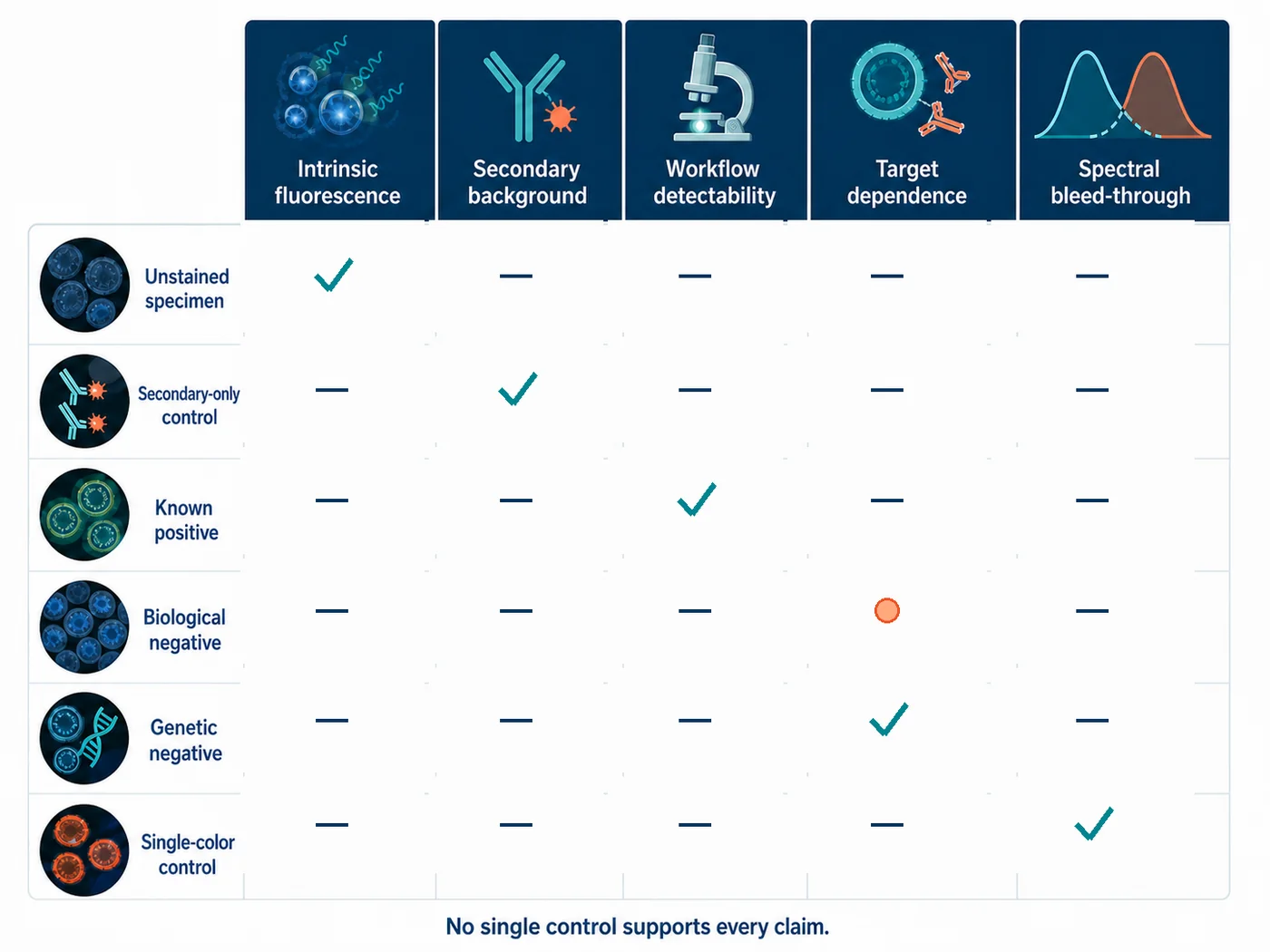
Choose controls for the conclusion you want to support
Different conclusions require different controls. Decide what the experiment must show before selecting the control set.
| Proposed conclusion | Main alternative explanation | Useful control or evidence |
|---|---|---|
| The target is detectable in this workflow. | The assay, retrieval, labeling, or acquisition failed. | Known-positive material processed in parallel, with expected morphology and localization. |
| The observed signal depends on the intended target. | The primary antibody recognizes an off-target structure or binds nonspecifically. | Application-matched genetic negative where feasible, supported by an independent antibody, orthogonal method, tagged-expression strategy, or other independent validation evidence. |
| The fluorescent secondary antibody is not creating the pattern. | The secondary antibody, endogenous immunoglobulin, Fc interaction, or detection reagent binds the specimen. | Primary-omission or secondary-only control that retains the complete downstream detection layer. |
| The specimen itself is not fluorescent in this channel. | Autofluorescence, fixative, pigment, substrate, mounting medium, or treatment produces signal. | Unstained or label-free specimen processed comparably and acquired with the same optical configuration. |
| A second channel represents a second fluorophore. | Cross-excitation, emission bleed-through, detector saturation, or spectral unmixing error contaminates the channel. | Single-color controls acquired through every channel using the final microscope configuration. |
| Two markers are present in the same cell or compartment. | Bleed-through, chromatic misregistration, out-of-focus light, segmentation error, or dense neighboring structures create apparent overlap. | Validated singleplex assays, single-color controls, individual-channel inspection, registration checks, and an analysis method matched to the biological scale of the claim. |
What each control can and cannot answer
| Control | What it checks | Limitation | When to include it |
|---|---|---|---|
| Unstained or label-free specimen | What intrinsic or preparation-related fluorescence is present in each channel? | Primary-antibody specificity or secondary-antibody background. | For each new specimen type, fixation, treatment, mounting condition, or unexpected background pattern. |
| Primary-omission / secondary-only | Does the downstream detection layer bind or fluoresce without the intended primary antibody? | Whether the primary antibody recognizes only the intended target. | With each experiment or labeling configuration that uses a secondary or amplification layer. |
| Known-positive material | Can the complete preparation, staining, and acquisition workflow detect the expected pattern? | Target dependence in the experimental sample. | With each run when failure would otherwise be indistinguishable from true absence. |
| Biological negative | Is signal lower in a specimen or compartment expected to lack or strongly reduce the target? | Complete molecular specificity or every source of background. | During assay establishment and when biological context changes. |
| Genetic negative | Does the staining pattern disappear or decrease when the target gene product is removed or strongly reduced? | That the genetic model is complete, free of residual protein, or biologically unchanged in all other respects. | During antibody characterization and major application changes; repeat when clone, preparation, or detection route changes materially. |
| Independent antibody | Do antibodies recognizing non-overlapping epitopes produce compatible target-dependent patterns? | Specificity when both reagents share the same off-target or when only display-level similarity is compared. | During validation of important or unexpected findings. |
| Single-color control | How much one fluorophore contributes to every other channel? | Antibody specificity, biological colocalization, or secondary background. | During panel design, after optical or fluorophore changes, and whenever contamination is suspected. |
| Batch reference | Did staining or acquisition performance change between runs? | Whether the assay is biologically specific. | Every batch used in longitudinal or quantitative comparisons. |
Controls for target dependence
Genetic controls
An isogenic knockout or well-characterized knockdown can provide strong application-specific evidence when the expected pattern is lost in the negative material and retained in the matched positive material. The genetic result must be interpreted with the preparation, antibody, and imaging settings used for immunofluorescence.
- Confirm that the model removes or substantially reduces the relevant protein or epitope, not merely its function.
- Use matched processing and acquisition for positive and negative material.
- Inspect all staining structures, not only the mean intensity.
- Consider residual protein, mosaic editing, alternative isoforms, compensatory biology, and altered morphology.
Independent-antibody and orthogonal evidence
Two antibodies recognizing non-overlapping epitopes can strengthen a conclusion when they show compatible, target-dependent patterns. Orthogonal evidence may include tagged-expression localization, RNA or protein abundance across a sample panel, biochemical fractionation, or another method with independent failure modes.
A western blot at the expected molecular mass does not establish that the same antibody produces a specific spatial pattern after fixation and permeabilization. Treat it as complementary evidence rather than a substitute for application-specific IF controls.
Known positive and known negative material
A useful positive control contains the target in a known anatomical, cellular, or subcellular location and is processed with the experimental specimens. A useful negative control lacks or strongly reduces the target under conditions that preserve enough comparable biology to make the contrast interpretable.
Controls for detection-reagent background
Indirect immunofluorescence
For a secondary-only control, omit the primary antibody but retain the same blocking, wash, fluorescent secondary antibody, counterstain, mounting, and acquisition steps. This tests the downstream detection layer under the actual experiment conditions.
In multiplex indirect IF, omission controls must match the panel design. Omitting one primary while retaining all secondaries can reveal whether a secondary recognizes the wrong primary, endogenous immunoglobulin, another detection antibody, or residual sites from a sequential step.
Directly conjugated primary antibodies
A conventional secondary-only control is irrelevant when no secondary is used. Background must instead be investigated with appropriate target-negative material, unstained specimens, reagent-omission controls, titration, Fc-blocking evaluation where relevant, and application-specific antibody validation.
Isotype and normal-immunoglobulin controls
An isotype-matched or species-matched nonimmune immunoglobulin can help diagnose Fc-receptor binding, reagent-class background, or concentration-dependent nonspecific interactions when it is matched as closely as practical for species, isotype, concentration, and conjugation.
A negative isotype result does not prove target specificity: the control and test antibodies have different variable regions, affinities, aggregation states, labeling ratios, and off-target interactions. Use it to investigate a defined reagent-class or Fc-binding mechanism, not as a universal substitute for target-negative evidence.
Controls for intrinsic fluorescence and imaging artifacts
Unstained or label-free specimen
Process a matched specimen through the relevant buffers, fixation, retrieval, treatment, mounting, and storage steps without fluorescent antibodies or dyes. Acquire it with the same objective, illumination, filter or detector bands, exposure logic, gain, and processing used for the stained sample.
Autofluorescence can be localized, particulate, diffuse, treatment-dependent, or channel-dependent. Increasing exposure until an unstained control becomes bright creates an acquisition artifact rather than additional biological information.
Single-color controls
Each fluorophore must be imaged alone through every channel in the final optical configuration. The control should match the specimen, fixation, mounting medium, labeling density, and acquisition method closely enough to reveal cross-excitation and emission bleed-through.
- Use raw channel values rather than merged display colors.
- Check contamination below saturation and at the exposure range used for the experiment.
- Repeat after changing fluorophore, filter set, laser line, detector band, objective, or unmixing method.
- Sequential acquisition can reduce some contamination but cannot correct fundamentally overlapping fluorophores or invalid controls.
Acquisition controls
Known-positive and control samples should establish channel identity, focus, specimen presence, detector response, saturation limits, and acceptable exposure before experimental images are interpreted. Quantitative comparisons require compatible acquisition settings, raw data retention, and documented processing.
Additional controls for multiplex IF
| Risk | Required control | What failure looks like |
|---|---|---|
| Spectral contamination | One single-color specimen for every fluorophore, acquired through all channels. | A fluorophore appears in a channel where it was not intended to be detected. |
| Secondary cross-detection | Omit each primary separately while retaining the complete secondary-antibody mixture. | A channel remains positive without its intended primary. |
| Same-host sequential labeling | Step-specific omission controls and tests for residual open binding sites. | A later primary or secondary is captured by an earlier detection layer, producing false overlap. |
| Panel interference | Compare every marker in final multiplex against its validated singleplex reference. | Distribution, intensity range, or background changes after other markers are added. |
| Batch drift | Stable reference material across staining and acquisition runs. | Reference intensity, morphology, or classification changes between batches. |
| False colocalization | Individual-channel review, registration check, z-position review, and validated segmentation. | Merged overlap disappears when optical, spatial, or analysis artifacts are corrected. |
See the Multiplex IF design guide for panel assembly, channel planning, acquisition, and panel checks.
Common control mistakes
| Mistake | Why the conclusion is invalid | Better interpretation |
|---|---|---|
| “No staining without primary antibody proves specificity.” | Primary omission mainly tests the secondary or downstream detection layer. | Report that secondary-related background was not detected under the tested conditions; use target-dependent evidence for primary-antibody specificity. |
| “The isotype control is negative, so the antibody is specific.” | The isotype reagent does not reproduce the test antibody’s variable region, affinity, aggregation, labeling ratio, or off-target binding. | Use it only to investigate a defined reagent-class or Fc-binding mechanism, alongside stronger target-dependent controls. |
| “Peptide absorption abolished staining, so the tissue pattern is specific.” | Absorption shows that the antibody binds the immunizing epitope; shared epitopes or multiple proteins may also be blocked. | Use absorption as supporting evidence and combine it with genetic, independent-antibody, or orthogonal validation. |
| “The western blot has one band, so the IF localization is validated.” | Denatured proteins on a membrane and fixed cellular structures expose different epitopes and have different off-target environments. | Treat western blotting as complementary evidence, then validate the immunofluorescence application directly. |
| “The signal matches the expected location, so it must be correct.” | Off-target proteins, autofluorescence, secondary binding, and processing artifacts can also produce plausible patterns. | Use expectation to define a testable pattern, not as evidence by itself. |
| “The strongest retrieval or antibody condition is best.” | Stronger signal can reflect increased background, tissue damage, saturation, or off-target accessibility. | Select conditions using positive and negative behavior, morphology, background, dynamic range, and reproducibility together. |
Practical minimum control sets
New single-target indirect IF assay
- Known-positive specimen or internal positive structure
- Target-negative biological or genetic material where feasible
- Primary-omission / secondary-only control
- Unstained or label-free specimen in every acquisition channel
- Primary and secondary antibody titration with acquisition below saturation
- Application-specific validation evidence recorded for the antibody
Established single-target assay used routinely
- Known-positive reference in the run
- Appropriate negative material or previously justified internal negative structure
- Secondary-only control when detection chemistry, specimen, lot, or background risk changes
- Unstained control when specimen type, fixation, treatment, mountant, or channel settings change
- Documented trigger for revalidation after clone, lot, preparation, detection, or instrument changes
Two-color or multicolor IF
- All controls required for every individual marker
- Single-color control for every fluorophore across all channels
- Method-matched omission controls for secondary or sequential cross-detection
- Singleplex-to-multiplex concordance for every marker
- Unstained specimen and stable batch reference
- Registration, saturation, and individual-channel review before merged interpretation
Plan the controls before staining
- State the biological conclusion you plan to make. Specify whether it concerns detectability, target dependence, spatial localization, abundance, co-expression, or colocalization.
- List the main alternative explanations. Include antibody off-target binding, secondary background, intrinsic fluorescence, spectral contamination, sample loss, acquisition error, and analysis error.
- Choose a control for each major alternative. Do not assign one control to questions it cannot answer.
- Define the expected result before staining. Record what should be present, absent, reduced, unchanged, or spatially restricted in each control.
- Process controls with the experiment. Match specimen preparation, incubation, washing, mounting, storage, and acquisition closely enough for the comparison to be meaningful.
- Acquire controls first. Confirm specimen presence, focus, channel identity, background, positive detectability, and exposure below saturation.
- Stop when a required control fails. Do not rescue an invalid run by adjusting display contrast or excluding inconvenient fields.
- Record when controls must be repeated. Decide which changes in antibody, lot, fluorophore, retrieval, specimen, instrument, or analysis require revalidation.
What to report
- Exact biological claim and unit of interpretation: specimen, region, cell, compartment, object, or pixel
- Specimen identity, preparation, fixation, permeabilization or retrieval, storage, and relevant treatment
- Primary antibody supplier, catalog number, clone, lot, host, isotype, concentration, dilution, and application-specific validation evidence
- Secondary or detection reagent identity, lot, cross-adsorption, fluorophore, concentration, dilution, and incubation
- Positive, negative, unstained, secondary-only, single-color, omission, singleplex-reference, and batch controls used
- Expected and observed outcome for every control, including failed or repeated controls
- Microscope, objective, illumination, filters or detector bands, exposure, gain, z-step, acquisition order, and saturation rule
- Raw-data retention, display adjustments, processing, registration, segmentation, thresholds, exclusions, and software version
- Changes that triggered revalidation and the evidence used to accept the revised assay
References and protocol sources
-
Richard W. Burry . Journal of Histochemistry & Cytochemistry (2011) . DOI: 10.1369/jhc.2010.956920
Functional separation of primary-antibody, secondary-antibody, and label controls; interpretation limits of omission and absorption controls.
- Controls for immunohistochemistry: The Histochemical Society’s standards of practice for validation of immunohistochemical assays Consensus guideline
Stephen M. Hewitt, Denis G. Baskin, Charles W. Frevert, William L. Stahl, and Eduardo Rosa-Molinar . Journal of Histochemistry & Cytochemistry (2014) . DOI: 10.1369/0022155414545224
Positive and negative control requirements, limits of primary-omission and absorption controls, and application-specific evidence for antibody specificity.
- A proposal for validation of antibodies Consensus guideline
Mathias Uhlén and colleagues . Nature Methods (2016) . DOI: 10.1038/nmeth.3995
Application-specific antibody validation using genetic, orthogonal, independent-antibody, tagged-expression, and capture-mass-spectrometry strategies.
- A consensus platform for antibody characterization Consensus guideline
Riham Ayoubi, Joel Ryan, Sara Gonzalez Bolivar, and colleagues . Nature Protocols (2025) . DOI: 10.1038/s41596-024-01095-8
Knockout-based, application-specific comparison of antibody performance in western blot, immunoprecipitation, and immunofluorescence.
- Immunofluorescence staining Methods paper
Julie G. Donaldson . Current Protocols in Cell Biology (2015) . DOI: 10.1002/0471143030.cb0403s69
Indirect immunofluorescence workflow, controls, fixation, permeabilization, and specimen handling.
- Bleed-through in fluorescence imaging Technical guide
Thermo Fisher Scientific
Recognition of signal from one fluorophore in a neighboring channel and mitigation through compatible fluorophore and filter selection.
- Selecting optical filters for fluorescence microscopy Technical guide
Thermo Fisher Scientific, Molecular Probes Handbook
Excitation filters, dichroic beamsplitters, emission filters, spectral separation, autofluorescence rejection, and instrument-aware optical trade-offs.
- The Society for Immunotherapy of Cancer statement on best practices for multiplex immunohistochemistry and immunofluorescence staining and validation Consensus guideline
Janis M. Taube and colleagues . Journal for ImmunoTherapy of Cancer (2020) . DOI: 10.1136/jitc-2019-000155
Singleplex-first panel development, marker-level controls, multiplex-to-singleplex concordance, and revalidation after panel changes.