Scope
This guide is intended for fixed cultured cells and tissue sections labeled with two or more fluorescent antibody channels. It covers conventional simultaneous indirect immunofluorescence, directly conjugated primary antibodies, and carefully validated mixed direct-and-indirect panels.
Cyclic staining, tyramide signal amplification, DNA-barcoded imaging, spectral pathology platforms, and other high-plex systems require platform-specific validation in addition to the principles below. A successful low-plex experiment does not establish that a cyclic or amplified version will preserve the same signal, morphology, or quantitative relationships.
Plan the panel around the biological question
| Check | Why it matters | Record |
|---|---|---|
| Required targets | Every additional marker adds antibody, spectral, control, and interpretation dependencies. | Target name, expected compartment, expected positive and negative material, and whether it is essential or optional. |
| Expected co-expression | Strongly co-expressed fluorophores are harder to separate than markers occupying different cells or compartments. | Expected cell types, compartments, relative abundance, and whether apparent overlap is biologically plausible. |
| Required conclusion | Detecting two signals in one field is different from proving cellular co-expression, subcellular colocalization, or molecular interaction. | Unit of analysis: field, tissue region, cell, compartment, object, or pixel; planned segmentation and exclusion rules. |
| Quantitative comparison | Quantitation requires compatible staining batches, acquisition settings, raw data, and analysis rules. | Comparison groups, batch design, reference material, acquisition order, saturation limit, and analysis pipeline. |
Choose the detection method
| Method | When it is useful | Main limitation |
|---|---|---|
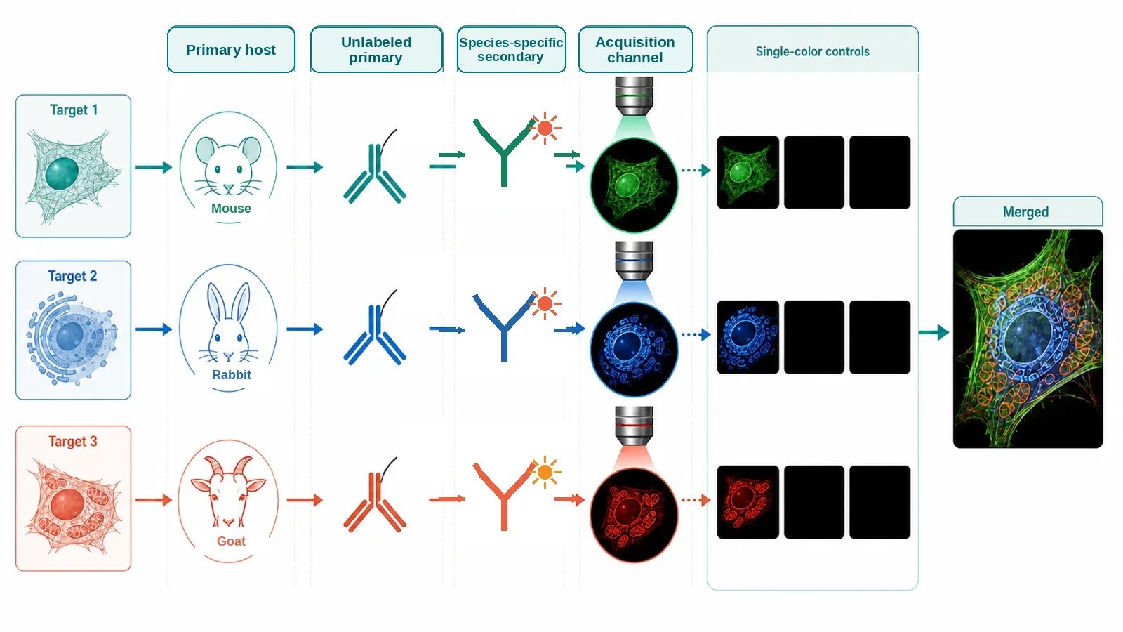
| Different-host unlabeled primaries with fluorescent secondaries | Simple low-plex panels when every primary host can be distinguished and the secondaries are compatible with the specimen. | Each secondary must recognize only its intended primary host. Cross-adsorption reduces unintended species reactivity but does not distinguish two primaries raised in the same host species. |
| Directly conjugated primary antibodies | Useful when host species overlap, when secondary amplification is unnecessary, or when reducing detection layers simplifies the panel. | Conjugation can change affinity or brightness, and each conjugated antibody requires its own titration and validation. |
| Mixed direct and indirect detection | Can preserve amplification for a dim target while using directly conjugated primaries for additional markers. | Order, open secondary-antibody binding sites, and cross-detection must be tested with omission controls. |
| Same-host unlabeled primaries with a validated sequential or Fab-blocking strategy | Specialized option when alternate host species or direct conjugates are not available. | Ordinary simultaneous secondary detection cannot identify which same-host primary generated a signal. Residual binding sites can create false overlap, so the complete sequence requires dedicated validation. |
| Cyclic, stripping, amplification, or barcoded platform | Higher-plex assays that exceed conventional simultaneous labeling. | Cycle order, target loss, incomplete stripping, registration, tissue damage, amplification, and platform-specific analysis introduce additional failure modes. |
When possible, use primary antibodies from different host species or validated directly conjugated primaries. Same-host workflows require additional controls because ordinary simultaneous secondary detection cannot distinguish the two primaries.
Validate every marker in singleplex first
- Confirm application suitability. Use an antibody supported for the relevant specimen preparation, fixation, retrieval, and species. Validation in western blotting or another assay does not establish immunofluorescence performance.
- Establish the expected pattern. Compare known-positive and target-negative material, cellular or tissue morphology, and the expected subcellular compartment.
- Titrate the primary and detection reagent. Select a condition that preserves the expected pattern and useful signal-to-background without detector saturation.
- Save the singleplex reference. Keep raw images, acquisition settings, representative fields, and control results for later comparison with the multiplex panel.
- Add markers one at a time. Do not move directly from single stains to the final panel.
- Compare multiplex with singleplex. Each marker should retain a comparable distribution and acceptable signal-to-background. Investigate a changed pattern rather than treating it as a display difference.
- Recheck the panel after material changes. Changing antibody clone, concentration, fluorophore, detection order, retrieval condition, or panel composition can alter other channels.
Plan fluorophores on the actual instrument
| Factor | Question | Check |
|---|---|---|
| Excitation | Does one excitation line substantially excite more than one fluorophore? | Excitation spectra, actual laser or illumination bands, and single-color images acquired in every channel. |
| Emission | Does one fluorophore emit into the detector or filter assigned to another channel? | Emission spectra, actual bandpasses, detector configuration, and measured bleed-through from single-color controls. |
| Specimen background | Which channels contain intrinsic fluorescence before antibodies are added? | Unstained specimen imaged with the same objective, filters, exposure logic, and processing used for the panel. |
| Target abundance | Which target is dim, abundant, diffuse, punctate, or expected to co-express strongly with another marker? | Singleplex intensity distribution below saturation, not supplier brightness rankings alone. |
| Optical sensitivity | Which channels have the best detector efficiency, transmission, illumination, and background on this microscope? | Instrument configuration and reference-sample measurements. |
| Photostability and order | Will early channels bleach while later channels are acquired? | Acquisition-order comparison or time-series check when exposure is substantial. |
Assigning a dim target to a favorable low-background channel can help, but there is no universal best color. The best channel depends on the fluorophore, labeling density, specimen, optics, detector, and exposure constraints.
Minimum control set
| Control | What it checks | How to use it |
|---|---|---|
| Unstained specimen | What intrinsic fluorescence is present in every acquisition channel? | Use the same specimen preparation and comparable acquisition settings as the multiplex sample. |
| Single-color control for every fluorophore | Does this fluorophore appear in another channel, and is a spectral reference required? | Acquire each single-color specimen across all channels. Match the fluorophore, specimen context, mounting medium, and acquisition configuration used in the panel. |
| Secondary-only or detection-reagent omission control | Does the detection layer create background without the intended primary antibody? | Match the omission control to the actual detection method; a directly conjugated panel does not use the same omission logic as an indirect panel. |
| Known-positive and target-negative material for each marker | Can the assay detect the expected marker, and does signal fall where the target is absent or strongly reduced? | Internal positive and negative structures are useful, but identify them explicitly rather than assuming them. |
| Singleplex reference for each marker | Does the multiplex panel reproduce the distribution obtained when the marker is stained alone? | Use compatible specimen preparation and acquisition; compare pattern and signal-to-background, not display colors alone. |
| Batch reference | Did staining or acquisition performance change between runs? | Include stable reference material when panels are run across days, batches, operators, or instruments. |
Panel assembly and staining
- Create a panel map before ordering reagents. List target, primary host species or isotype, antibody clone, detection route, fluorophore, expected abundance, expected compartment, and planned controls.
- Keep specimen preparation fixed. Use the fixation, permeabilization, or retrieval condition established during singleplex validation. Do not change specimen preparation and multiplex chemistry at the same time unless the comparison is explicitly designed.
- Prepare all controls in the same run. Include unstained, single-color, appropriate omission, positive, negative, and batch-reference specimens before beginning the multiplex sample.
- Block according to the chosen detection method. Consider specimen immunoglobulins, secondary-antibody hosts, Fc interactions, and any Fab-blocking or sequential strategy.
- Apply compatible primary antibodies. Simultaneous incubation is reasonable only when the detection layers can distinguish every primary. Otherwise use a validated direct, sequential, or platform-specific route.
- Wash consistently. Keep buffer, volume, duration, count, and agitation consistent across multiplex and control specimens.
- Apply fluorescent detection. For indirect panels, use species-appropriate and sufficiently cross-adsorbed secondaries. For directly conjugated panels, protect from light and preserve the validated conjugate concentration.
- Complete any validated blocking or sequential step. Confirm that residual detection sites cannot capture a later primary or conjugate. Include omission controls that isolate each step.
- Counterstain and mount consistently. Avoid a counterstain whose spectrum compromises a required marker. Keep mountant, coverslip, curing, and storage interval consistent.
- Acquire the controls before interpreting the panel. Confirm specimen background, channel contamination, expected positive signal, and negative-control behavior before drawing conclusions from the multiplex image.
Acquisition workflow
- Acquire the unstained specimen first. Measure baseline fluorescence in every planned channel and note tissue-, treatment-, substrate-, or fixative-related patterns.
- Acquire every single-color control across all channels. Measure cross-excitation and emission bleed-through using the actual optical configuration.
- Use sequential acquisition when appropriate. Exciting and detecting one fluorophore at a time can reduce cross-excitation and bleed-through, but it does not correct poor fluorophore selection or invalid controls.
- Set exposure below saturation. Use the positive reference and brightest expected specimen to identify a usable range. Preserve the same acquisition logic across groups intended for quantitative comparison.
- Record channel order and illumination history. Fixed specimens can still bleach, and acquisition order can alter later channels.
- Save raw individual channels. Preserve raw data and metadata before flat-field correction, deconvolution, spectral unmixing, denoising, thresholding, segmentation, or display adjustment.
- Validate spectral unmixing when used. Reference spectra must match the fluorophore, specimen background, and acquisition configuration closely enough for the chosen method.
- Inspect channels individually before merging. A visually plausible merged image can conceal saturation, bleed-through, misregistration, background, or a failed channel.
Before using the panel for experiments
- Every marker has an acceptable singleplex assay with an expected pattern, known-positive behavior, and target-negative evidence.
- Each marker retains a comparable distribution and acceptable signal-to-background in the final multiplex panel.
- Single-color controls show that measured channel contamination is absent, acceptable, or handled by a validated acquisition and unmixing method.
- Unstained and detection-reagent controls define intrinsic and detection-layer background in every relevant channel.
- No required marker is saturated in specimens used for quantitative comparison.
- Panel performance is reproducible across the required days, batches, operators, and reference specimens.
- Tissue or cellular morphology remains adequate for the intended segmentation and biological interpretation.
- Recheck affected markers after changing the clone, concentration, fluorophore, staining order, retrieval condition, or panel composition.
Interpretation limits
- Pixel overlap is not molecular interaction. Apparent colocalization can arise from diffraction, out-of-focus light, channel bleed-through, chromatic misregistration, dense structures, or segmentation error.
- A merged display is not raw evidence. Conclusions should be traceable to the individual channels, controls, acquisition metadata, and analysis rules.
- Display balance can mislead. Independent contrast scaling can make weak contamination look equivalent to strong target signal. Preserve raw values and disclose display transformations.
- Segmentation and phenotyping require validation. Inspect errors across representative tissue regions and biological conditions rather than validating an algorithm on a few attractive fields.
- Batch correction cannot rescue an invalid assay. Computational normalization does not replace marker-level controls, stable reference material, and reproducible staining.
Troubleshooting checkpoints
| Observation | Check first | Next action |
|---|---|---|
| Signal appears in two channels | Single-color control acquired across both channels, excitation sequence, emission filters, and saturation. | Separate fluorophores or optical bands, use sequential acquisition, reduce saturation, or validate spectral unmixing. |
| Two same-host markers overlap unexpectedly | Omission controls for each primary, residual secondary binding sites, blocking order, and direct-conjugate detection. | Redesign with different hosts or direct conjugates, or validate a Fab-based or sequential strategy independently. |
| A marker works alone but fails in multiplex | Primary and secondary competition, altered concentration, staining order, retrieval, fluorophore brightness, and exposure. | Add markers incrementally and compare each stage with the singleplex reference. |
| Multiplex pattern differs from singleplex | Clone, fluorophore, detection chemistry, order, amplification, registration, and analysis thresholds. | Do not use the panel until the cause is identified and the marker is rechecked. |
| Dim target is lost | Singleplex signal, specimen background, channel sensitivity, fluorophore assignment, detector saturation elsewhere, and antibody competition. | Use a more favorable measured channel or validated amplification route without changing multiple variables at once. |
| Merged image suggests false colocalization | Individual channels, single-color controls, chromatic registration, z-position, segmentation, and optical resolution. | Correct the optical or analysis cause and report only the level of co-occurrence supported by the data. |
| Results drift between batches | Reference specimen, reagent lots, incubation timing, temperature, instrument calibration, acquisition settings, and analysis version. | Identify the affected stage before applying batch correction or combining data. |
What to record for a multiplex panel
- Biological question, specimen type, comparison groups, target list, expected compartments, and expected co-expression
- Specimen preparation, fixation, permeabilization, retrieval, section thickness, slide or imaging surface, and storage history
- Primary antibody supplier, catalog number, clone, lot, host species, isotype, concentration, dilution, and singleplex validation evidence
- Detection route for every marker: direct conjugate, secondary antibody, amplification, sequential step, Fab block, or platform cycle
- Secondary or detection reagent identity, lot, cross-adsorption, concentration, fluorophore, incubation order, and blocking sequence
- Instrument lasers or illumination bands, dichroics, emission filters, detectors, objective, exposure, gain, binning, z-step, acquisition order, and saturation rule
- Unstained, single-color, omission, positive, negative, singleplex-reference, and batch-reference controls
- Panel-check results for every marker and reason for any deviation from the singleplex reference
- Raw-file location, metadata, flat-field correction, deconvolution, unmixing, registration, segmentation, thresholds, phenotype rules, exclusions, and software version
- Batch, operator, date, reagent-lot changes, instrument changes, revalidation decisions, and representative uncropped control images
References and protocol sources
- The Society for Immunotherapy of Cancer statement on best practices for multiplex immunohistochemistry and immunofluorescence staining and validation Consensus guideline
Janis M. Taube and colleagues . Journal for ImmunoTherapy of Cancer (2020) . DOI: 10.1136/jitc-2019-000155
Singleplex-first panel development, marker-level controls, multiplex-to-singleplex concordance, and revalidation after panel changes.
- Multiplexed IHC staining with primary antibodies conjugated to fluorophores Manufacturer protocol
Thermo Fisher Scientific
Directly conjugated primary-antibody workflow, single-color controls, negative controls, coverage, and antibody titration.
- Labeling primary antibodies from the same host species Manufacturer protocol
Jackson ImmunoResearch
Why ordinary secondary detection cannot distinguish same-host unlabeled primaries and how Fab-based or mixed direct/indirect strategies can be designed.
- Selecting optical filters for fluorescence microscopy Technical guide
Thermo Fisher Scientific, Molecular Probes Handbook
Excitation filters, dichroic beamsplitters, emission filters, spectral separation, autofluorescence rejection, and instrument-aware optical trade-offs.
- Guide for using the Fluorescence SpectraViewer Technical guide
Thermo Fisher Scientific
Instrument-aware fluorophore compatibility and quantitative inspection of spectral overlap.
- Society for Immunotherapy of Cancer: updates and best practices for multiplex immunohistochemistry and immunofluorescence image analysis and data sharing Consensus guideline
Janis M. Taube and colleagues . Journal for ImmunoTherapy of Cancer (2025) . DOI: 10.1136/jitc-2024-008875
Acquisition, segmentation, phenotyping, quality control, batch correction, and sharing of raw and processed outputs.
- Society for Immunotherapy of Cancer: Standards for Reporting of Multiplex Immunohistochemistry/Immunofluorescence Assays (STORMI) Consensus guideline
Sam Sater and colleagues . Journal for ImmunoTherapy of Cancer (2025) . DOI: 10.1136/jitc-2025-012280
Minimum reporting elements for panel design, staining controls, acquisition, analysis, and reproducibility.