Scope
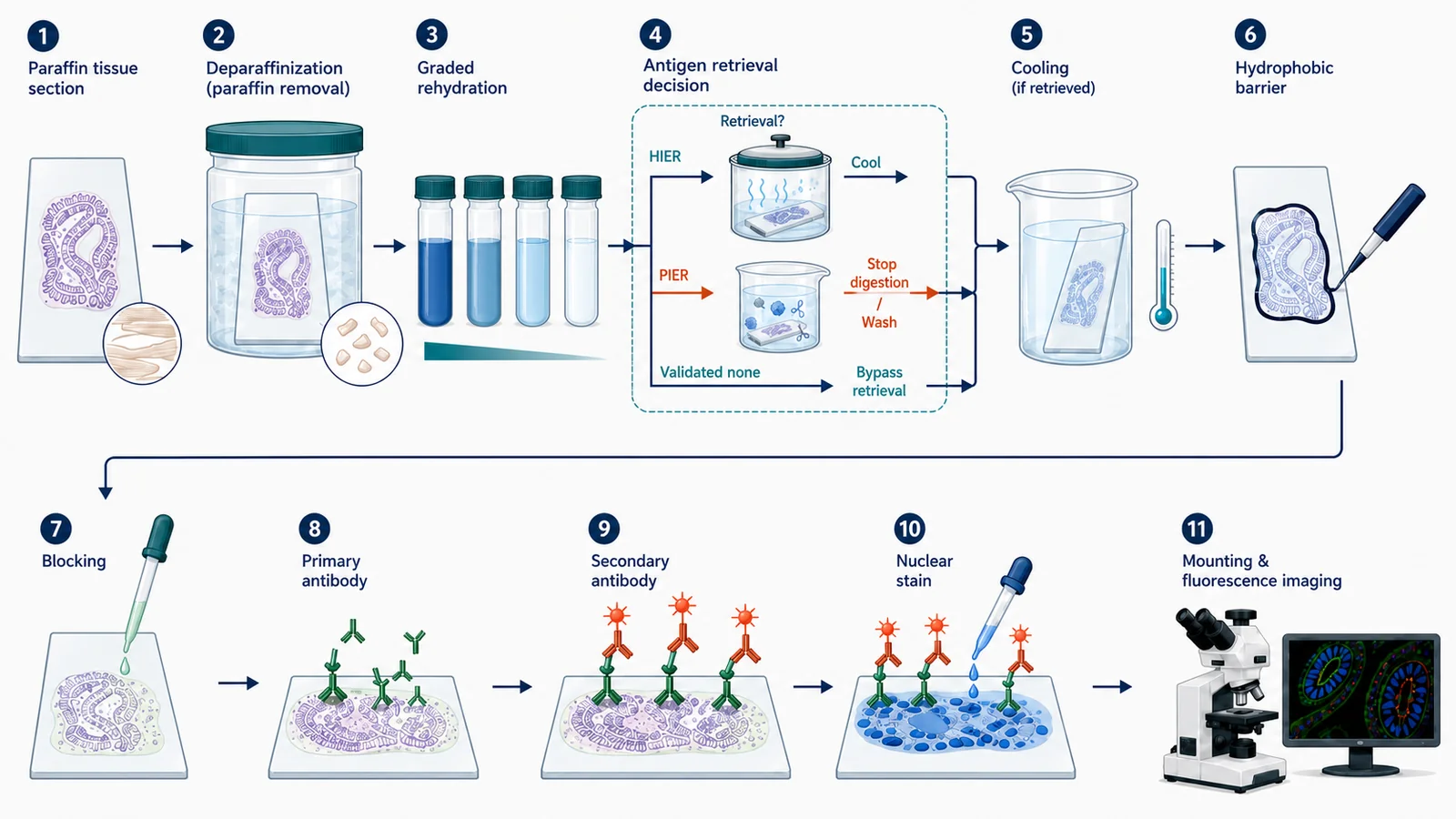
This protocol is intended for formalin-fixed paraffin-embedded tissue sections mounted on microscope slides and detected by direct or indirect fluorescence. The workflow covers paraffin removal, rehydration, antigen retrieval, staining, and imaging.
An antibody validated for cultured cells or frozen tissue should not be assumed to work in FFPE. Antibodies validated for chromogenic IHC-P may sometimes support fluorescent detection, but target abundance, fixation history, autofluorescence, and amplification requirements must be assessed separately.
Before you start
| Check | Why it matters | Record |
|---|---|---|
| Pre-analytical tissue history | Cold ischemia, fixative type, fixation duration, processing temperature, paraffin infiltration, and storage can alter epitope accessibility and background. | Collection delay, tissue size, fixative, fixation duration, processor program, embedding date, block age, and storage conditions. |
| Section quality | Thickness, folds, chatter, incomplete adhesion, and compression change reagent access and apparent signal density. | Nominal thickness, slide type, baking conditions, orientation, folds, detachment, and tissue loss. |
| Antibody validation | Validation in another application does not establish performance after formalin fixation and paraffin embedding. | IHC-P or FFPE validation evidence, expected compartment and morphology, target abundance, and known-positive tissue. |
| Retrieval method | Heat and enzymes may increase target access but can also increase background, detach tissue, or damage morphology. | Buffer identity, pH, device, temperature profile, duration, cooling, enzyme concentration, and stop method. |
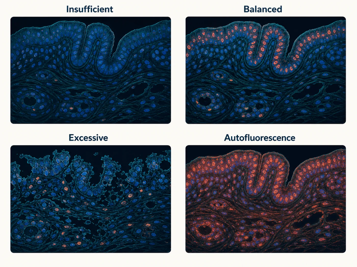
| Baseline fluorescence | Formalin-fixed tissue, pigments, lipofuscin, red blood cells, collagen, and treatment history can contribute channel-dependent fluorescence. | Unstained section acquired in every planned channel with the same objective and comparable settings. |
| Imaging and amplification | Low-abundance targets may not be detectable by simple indirect fluorescence, while amplification can narrow dynamic range or increase background. | Detection chemistry, fluorophores, microscope, channels, exposure, gain, z-step, saturation limit, and processing rules. |
Materials and reagents
| Category | Required | Selection notes |
|---|---|---|
| Sections and slides | FFPE tissue sections on positively charged or otherwise validated adhesive slides | Use a slide type and baking condition proven to retain the tissue during the intended retrieval method. |
| Clearing and rehydration | Xylene, xylene substitute, or validated clearing agent; graded histology-grade ethanol; purified water | Use fresh, uncontaminated solutions and a documented sequence. Incomplete paraffin removal can produce patchy access and poor wetting. |
| Retrieval | Validated low-pH, neutral, or high-pH HIER buffer; or a validated protease for PIER | Choose from antibody and tissue evidence. Do not combine heat and enzyme retrieval unless deliberately validated. |
| Wash buffer | PBS or TBS | Use one defined formulation consistently across comparison groups. |
| Blocking and antibody diluent | Normal serum, BSA, or validated commercial buffer | Match normal serum to the secondary-antibody host when appropriate and avoid components that interfere with the detection chemistry. |
| Primary antibody | Antibody validated for FFPE/IHC-P or supported by local FFPE evidence | Record supplier, catalog number, clone, lot, concentration, dilution, and validation application. |
| Detection | Fluorophore-conjugated primary, species-specific fluorescent secondary, or validated amplification chemistry | Highly cross-adsorbed secondaries are preferred for multiplex work. Confirm that amplification is necessary before adding complexity. |
| Counterstain and mounting | Nuclear stain as needed, compatible antifade mounting medium, coverslip | Choose spectra and mountant compatible with the instrument, tissue background, and planned storage. |
| Handling | Slide rack or jars, humidified chamber, hydrophobic barrier pen if compatible, light protection | Use enough volume to cover the complete section and prevent drying after rehydration. |
Common starting conditions
These starting conditions are taken from current manufacturer protocols. Product-specific instructions and direct comparisons should determine the final condition for each antibody and tissue.
| Stage | Starting condition | Check |
|---|---|---|
| Slide warming | Approximately 20 minutes at 50–60°C | Can improve adhesion and soften paraffin. Avoid exceeding the validated temperature because excessive heat can damage some targets. |
| Deparaffinization and rehydration | Example sequence: clearing agent 3 × 5 minutes; 100% ethanol 2 × 5 minutes; 90% ethanol 5 minutes; 70% ethanol 5 minutes; purified water 5 minutes | A documented starting sequence used in current Thermo Fisher protocols. Equivalent validated sequences may be used, but incomplete deparaffinization must be ruled out. |
| High-pH HIER | Example: fully submerged slides at approximately 98°C for 20 minutes, followed by cooling in retrieval solution for about 20 minutes | A product-specific starting point, not a universal FFPE recipe. Compare morphology, signal, background, and tissue retention. |
| Proteolytic retrieval | Example: 0.1% trypsin for approximately 10–20 minutes at 37°C | Time and enzyme concentration are tissue- and antigen-dependent. Stop digestion and wash promptly to avoid morphology loss. |
| Blocking | Approximately 60 minutes at room temperature | Evaluate with the secondary-only control rather than assuming longer blocking always improves specificity. |
| Primary antibody | Manufacturer-recommended dilution, commonly overnight at 4°C | Titrate concentration and compare known-positive tissue, target-negative tissue, localization, and background. |
| Secondary antibody | Manufacturer-recommended dilution for approximately 1 hour at room temperature, protected from light | Match the primary host species and include a secondary-only section. |
| Washing | Three gentle washes of approximately 5 minutes between major labeling steps | Keep composition, duration, volume, and agitation consistent while protecting tissue adhesion. |
Procedure
- Document the block and section. Record tissue history, fixation, processing, block age, section thickness, slide type, orientation, folds, compression, and adhesion before staining.
- Warm the slide when required. Use the validated temperature and duration needed for adhesion and paraffin softening. Do not use excessive heat as a substitute for an inadequate slide or sectioning method.
- Remove paraffin completely. Immerse slides in fresh clearing reagent using a documented sequence. Agitate gently enough to dislodge bubbles and improve solvent exchange without damaging the section.
- Rehydrate through graded alcohols. Move from high to lower alcohol concentration and then to water or buffer using defined times. From the final aqueous step onward, do not allow the section to dry.
- Apply the selected retrieval condition. For HIER, keep the tissue fully submerged, control the actual solution temperature, and use a reproducible cooling phase. For PIER, control enzyme identity, concentration, temperature, duration, and stop method. Skip retrieval only when the antibody has been shown to work without it in the relevant preparation.
- Wash and inspect tissue retention. Before using antibody, confirm that the section remains attached and that morphology is adequate. Record folds, cracks, edge lifting, or tissue loss caused by retrieval.
- Mark the staining area and measure background. Apply a compatible barrier after the slide perimeter is dry enough for the pen while keeping tissue hydrated. Acquire an unstained serial section in every planned channel.
- Block nonspecific interactions. Cover the tissue completely in a humidified chamber. Prepare antibody dilutions during this period using one defined diluent for comparable sections.
- Incubate the primary antibody. Record antibody identity, clone, lot, concentration, dilution, diluent, time, temperature, volume, and whether amplification will be used.
- Wash consistently. Use gentle immersion or low-force exchange with the same number, duration, volume, and agitation for every comparable section.
- Apply fluorescent detection. Use a species-appropriate fluorescent secondary, directly conjugated primary, or validated amplification method. Protect from light and prevent cross-reactivity in multiplex panels.
- Wash, counterstain, and mount. Remove unbound fluorophore, choose a non-overlapping counterstain, prevent bubbles and compression, and use a compatible antifade mountant.
- Acquire controls before test sections. Confirm tissue presence, intrinsic fluorescence, secondary background, known-positive signal, channel identity, and exposure below saturation before imaging experimental groups.
Minimum control set
| Control | What it checks | Limitation |
|---|---|---|
| Unstained serial section | How much tissue-, pigment-, formalin-, treatment-, and channel-related fluorescence is present without antibody? | Antibody specificity or secondary-antibody background. |
| Secondary-only section | Does the fluorescent secondary antibody or detection chemistry create background without the primary antibody? | That the primary antibody recognizes only the intended target. |
| Known-positive FFPE tissue | Can this fixation, retrieval, staining, and acquisition workflow detect the expected pattern? | Target dependence in the experimental tissue. |
| Biological or genetic negative | Does signal decrease where the target is absent or substantially reduced? | All sources of intrinsic fluorescence, bleed-through, or secondary background. |
| Retrieval comparison | Does changing pH, heat, enzyme, or retrieval omission alter signal and morphology in a target-consistent way? | That the strongest signal is the most specific or least damaged condition. |
| Single-color control | Does one fluorophore appear in another acquisition channel? | Target specificity or nonspecific antibody binding. |
For low-abundance FFPE targets, a failed indirect fluorescent readout does not automatically mean the target is absent. First confirm antibody suitability, positive-control performance, retrieval, intrinsic fluorescence, and whether an amplification strategy is justified.
Stop points and storage
- Paraffin blocks: store according to laboratory policy and record block age, storage conditions, and recutting history.
- Unstained sections: storage duration can change adhesion and antigenicity. Use a validated schedule and record sectioning date, slide type, and storage environment.
- After deparaffinization and rehydration: do not allow tissue to dry. Continue in aqueous buffer or retrieval solution according to a controlled schedule.
- After retrieval: complete the specified cooling or enzyme-stop step, then proceed without uncontrolled drying or prolonged delay.
- During primary incubation: overnight incubation at 4°C is a planned step. Maintain humidity and complete tissue coverage.
- After mounting: allow the mountant to cure as directed, then store flat at 4°C protected from light and record the interval before imaging.
Troubleshooting checkpoints
| Observation | Check first | Next guide |
|---|---|---|
| No or weak target signal | Known-positive FFPE tissue, antibody validation application, tissue presence, acquisition settings, paraffin removal, and retrieval condition. | No-signal workflow |
| High diffuse background | Secondary-only section, exposure saturation, antibody titration, wash consistency, tissue drying, amplification strength, and barrier compatibility. | High-background patterns |
| Strong channel-wide fluorescence | Unstained serial section in every channel, formalin history, endogenous pigments, red blood cells, collagen, lipofuscin, treatment, and mounting medium. | Autofluorescence by channel |
| Patchy or hydrophobic-looking staining | Incomplete paraffin removal, bubbles, inadequate rehydration, tissue drying, incomplete coverage, and local detachment. | Uneven-staining patterns |
| Section lifting after retrieval | Slide adhesion, baking, section age, retrieval pH and heat, pressure changes, wash force, and tissue composition. | Uneven-staining patterns |
| Strong signal with damaged morphology | Retrieval temperature, time, pH, enzyme concentration, cooling, fixation duration, and section thickness. | Antigen-retrieval guide |
| Low-abundance target not detectable | Known-positive signal, antibody suitability for FFPE, fluorophore brightness, intrinsic fluorescence, optical settings, and need for validated amplification. | Fluorophore-selection guide |
What to record
- Tissue identity, source, biological replicate, collection delay, fixative, fixation duration, and tissue dimensions
- Processor program, dehydration and clearing history, paraffin, embedding date, block age, and storage
- Section thickness, slide type, sectioning date, baking conditions, folds, compression, and adhesion
- Clearing reagent, batch or age, deparaffinization and graded-rehydration sequence
- Retrieval buffer or enzyme, pH, concentration, device, actual temperature, duration, cooling or stop method, and tissue-retention result
- Blocking, primary, secondary, and amplification reagents with identifiers, lots, concentrations, dilutions, times, temperatures, and volumes
- Wash composition, count, duration, volume, agitation, and any section lifting
- Unstained, secondary-only, positive, negative, retrieval-comparison, and single-color controls as applicable
- Mounting medium, coverslip, curing and storage interval, microscope, objective, channels, exposure, gain, z-step, and processing
- Raw control images, uncropped representative fields, excluded sections, and reasons for exclusion
References and protocol sources
- FFPE tissue high-pH antigen retrieval, direct fluorescent method Manufacturer protocol
Thermo Fisher Scientific
Slide warming, deparaffinization, graded rehydration, high-pH HIER, cooling, staining, and storage.
- FFPE tissue trypsin digestion antigen retrieval, indirect fluorescent method Manufacturer protocol
Thermo Fisher Scientific
Proteolytic retrieval starting conditions and indirect fluorescent detection workflow.
- Paraffin removal and antigen retrieval Technical guide
Thermo Fisher Scientific
Deparaffinization, rehydration, and epitope retrieval as distinct preparation stages.
-
Cell Signaling Technology
FFPE application validation, formalin-associated autofluorescence, sensitivity limits, and amplification considerations.
- IHC antigen retrieval protocol Manufacturer protocol
Abcam
Heat-induced and enzymatic retrieval as target- and tissue-dependent strategies.