Scope
This guide is for fixed-cell and tissue immunofluorescence in which the expected target signal is absent or weaker than required for interpretation. It covers cultured cells, frozen sections, FFPE sections, direct and indirect detection, and low-plex multiplex panels.
“No detectable signal” is an observation, not proof that the target is absent. Do not conclude that the target is absent until specimen presence, positive-control performance, target-dependent controls, sample preparation, detection chemistry, and acquisition have been checked.
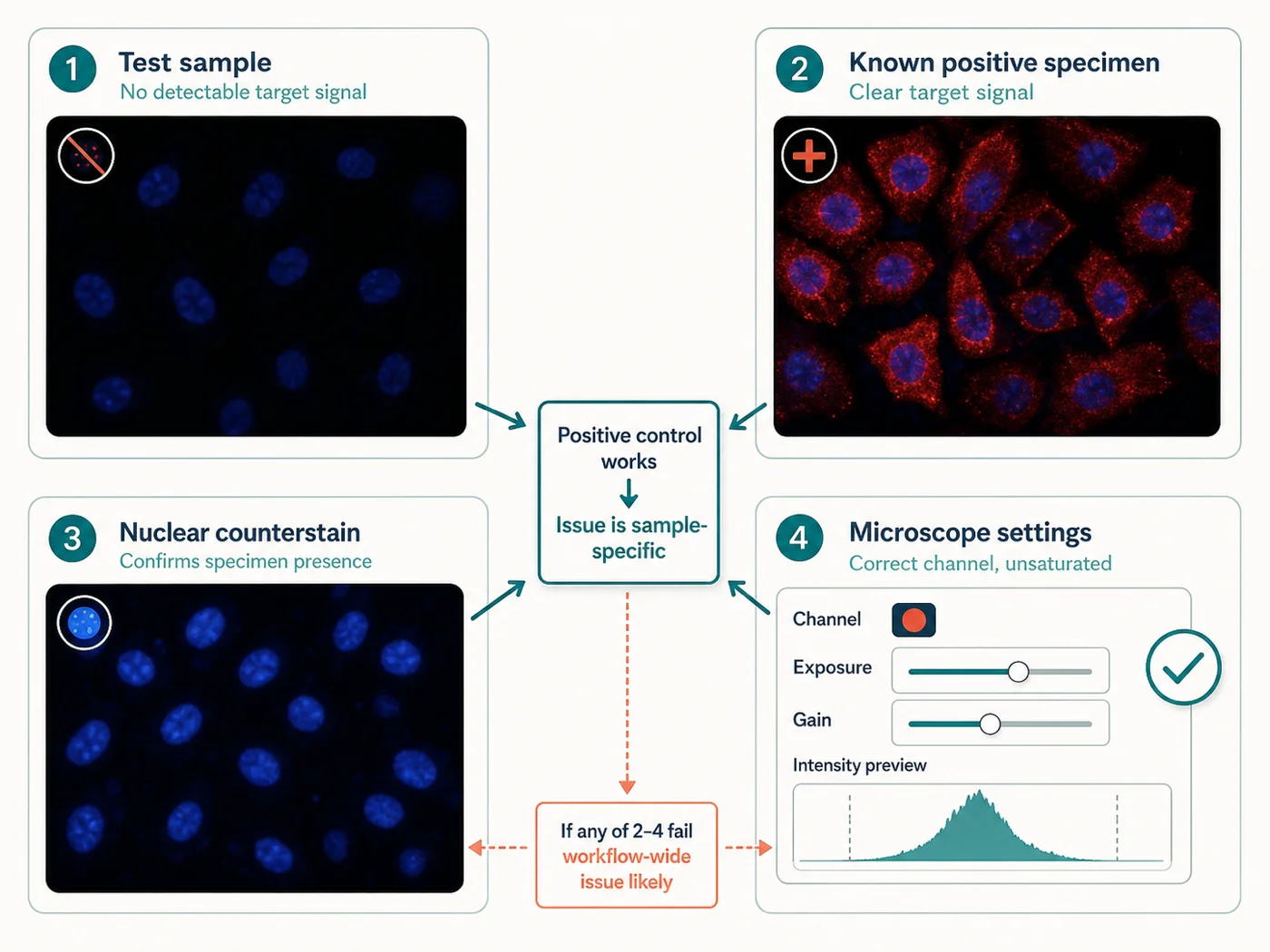
Quick check: where did the signal fail?
| Observation | Likely explanation | Check next |
|---|---|---|
| Specimen is not visible by transmitted light, morphology channel, or counterstain | Sample loss, wrong focal plane, mounting failure, field-selection error, or acquisition problem is more likely than antibody failure | Inspect the slide, coverslip, tissue boundary, brightfield or transmitted light, and a validated structural or nuclear reference |
| Known-positive control also has no target signal | A problem involving reagent identity, detection method, preparation, or acquisition is more likely | Confirm antibody and secondary identities, fluorophore, channel, sequence, reagent age, and a previously successful preparation |
| Known-positive control works, but test sample fails | Biological absence, low abundance, sample-specific handling, treatment effect, or preparation-specific masking becomes more plausible | Check sample history, independent expression evidence, target-negative material, and an orthogonal or independent-antibody result |
| Signal is present in singleplex but absent after multiplex assembly | Panel interference, host-species incompatibility, fluorophore reassignment, order effect, spectral contamination, or cumulative treatment is more likely | Compare the singleplex reference with the multiplex channel under matched acquisition and omission controls |
| Signal appears only at extreme exposure or gain | Low signal-to-background, poor optical compatibility, weak labeling, photobleaching, or true low abundance remains unresolved | Inspect raw values, unstained background, positive control, single-color control, channel assignment, and saturation elsewhere in the field |
| Only one region or edge lacks signal | Coverage, drying, bubbles, section detachment, illumination, focus, or specimen thickness is more likely than global reagent failure | Inspect the physical pattern before changing antibody or retrieval |
Step 1: confirm that the specimen is present
Do not troubleshoot antibody concentration until the sample itself is confirmed.
- Locate the complete cell field or tissue boundary using transmitted light, a morphology reference, or a validated counterstain
- Confirm that cells did not detach during washes and that the tissue section did not lift, fold, digest, or wash away
- Check the coverslip, mounting medium, orientation, bubbles, compression, and whether the specimen faces the objective
- Verify the focal plane and z-range rather than relying on one position
- Confirm that the field-selection method did not choose an empty, necrotic, damaged, or biologically irrelevant region
- Inspect whether local drying, hydrophobic barriers, incomplete reagent coverage, or bubbles excluded part of the specimen
Step 2: check the microscope and acquisition settings
| Check | Failure that can mimic no signal | How to check it |
|---|---|---|
| Excitation source | The selected laser, LED, or excitation filter does not efficiently excite the fluorophore | Instrument configuration and fluorophore spectrum, followed by a known fluorescent control |
| Emission path | The emission filter or detector window excludes much of the fluorophore signal | Actual bandpass or spectral window and a single-color control acquired through the final channel |
| Detector and settings | Wrong detector, closed shutter, low gain, short exposure, incorrect binning, or disabled channel | Saved acquisition settings and a control known to emit in that channel |
| Focus and z-position | The target lies outside the acquired focal plane or section thickness | Focus search using morphology, then a limited z-stack where appropriate |
| Sequential or simultaneous setup | Wrong sequence, inactive channel, mismatched filter wheel, or spectral-unmixing reference | Raw single-color control through every final acquisition channel |
| Display range | Signal exists in raw data but is hidden by display scaling, incorrect lookup table, or background subtraction | Raw intensity histogram and image before processing |
| Photobleaching | Signal was lost during focusing, repeated scanning, long z-stacks, or delayed acquisition | Compare the first and last frames, reduce illumination, and acquire a fresh field |
Do not use extreme exposure as the first diagnostic. It may reveal background, bleed-through, or detector noise rather than target-dependent signal.
Step 3: check the positive control
| Positive-control result | What this result means | Next action |
|---|---|---|
| Positive control fails under the same preparation and acquisition | Do not interpret the test sample as negative | Check reagent identities, preparation, staining order, detection compatibility, storage, and acquisition before changing the biological sample |
| Positive control works with the same antibody and detection layer | The workflow can detect the target in at least one context | Investigate test-sample expression, handling, fixation, retrieval, treatment timing, target abundance, and specimen integrity |
| Positive control works only under a different preparation | Antibody performance has not been established for the failed application | Use application-specific evidence or compare preparation routes while keeping antibody and acquisition constant |
| Positive control is only a highly overexpressing model | Detectability in the test sample may still be inadequate | Add a positive material closer to the expected abundance and specimen type |
| Positive control is old, poorly documented, or lacks expected localization | It is not a reliable workflow reference | Re-establish a traceable positive control with expected compartment and morphology |
A positive control should show whether the actual antibody, preparation, detection chemistry, and microscope can produce the expected spatial pattern. Fluorescence somewhere in the specimen is not enough.
Step 4: check the antibody and detection reagents
| Detection method | Common no-signal cause | Check |
|---|---|---|
| Indirect IF | Secondary antibody does not recognize the primary host species or immunoglobulin class | Primary host, isotype, secondary specificity, conjugate, and species compatibility |
| Directly conjugated primary | Conjugation reduced binding, fluorophore faded, or the degree of labeling is unsuitable | Unconjugated or previously validated equivalent, conjugate lot, storage, and positive control |
| Multiple primaries and secondaries | Host-species conflict, missing secondary, cross-adsorption assumptions, or sequential-layer blocking error | One-marker-at-a-time controls and method-matched omission controls |
| Amplified detection | Missing activation step, incompatible buffer, incorrect sequence, or exhausted reagent | Complete manufacturer-validated sequence and a positive amplification control |
| Spectral or cyclic workflow | Wrong fluorophore cycle, stripping damage, mismatched reference spectrum, or cumulative target loss | Cycle-specific controls, singleplex reference, and raw data from the failed cycle |
- Confirm supplier, catalog number, clone, lot, host, isotype, conjugate, fluorophore, concentration, dilution, and expiration or preparation date
- Confirm that the correct reagent was placed in the correct tube, well, slide, or staining step
- Check whether light-sensitive reagents were protected and whether repeated freeze-thaw, precipitation, or contamination occurred
- Confirm that incubation volume fully covered the specimen and that humidity prevented evaporation
- Verify the wash sequence did not accidentally omit a reagent or remove a loosely attached specimen
Step 5: check whether the target should be present
| Biological question | Why signal may be weak or absent | Useful evidence |
|---|---|---|
| Is the target expressed in this sample? | Cell type, tissue region, differentiation state, treatment, disease state, passage, or donor may differ from the expected context | Independent expression data from a matched specimen and known target-negative material |
| Was the target present at the sampled time? | Transient induction, degradation, trafficking, phosphorylation, secretion, or internalization may shift rapidly | Time-course evidence and a positive control processed at the same time |
| Is the expected compartment correct? | The target may move between nucleus, cytoplasm, membrane, organelle, extracellular space, or a soluble pool | Singleplex localization, compartment markers, and an independent antibody or tagged model when justified |
| Is abundance below assay sensitivity? | Valid target may be present but below the signal-to-background achievable with the current labeling and instrument | Known-positive material near the expected abundance, a more favorable channel, and an orthogonal method |
| Does the antibody recognize the relevant form? | Isoform, cleavage, modification, conformation, species, or epitope accessibility may differ | Antibody documentation, genetic negative, independent antibody, or orthogonal validation |
Western blotting, RNA measurements, public expression databases, or overexpression can support a hypothesis but do not alone validate the spatial pattern in the fixed specimen. Antibody validation remains application-specific.
Step 6: change one sample-preparation factor at a time
| Preparation step | How it can suppress signal | Comparison to try |
|---|---|---|
| Fixation | Overfixation, delayed fixation, solvent extraction, redistribution, or epitope masking | Compare one validated aldehyde route with one antibody-supported alternative while holding labeling and imaging constant |
| Permeabilization | Insufficient access to the required compartment or excessive extraction of the target | Compare no detergent with one mild validated condition; add a stronger condition only when justified |
| FFPE deparaffinization and rehydration | Residual paraffin or drying prevents reproducible wetting and access | Repeat with a documented clearing and rehydration sequence before changing retrieval |
| Antigen retrieval | Inadequate route leaves the epitope inaccessible; excessive heat or protease destroys tissue or target | Compare validated no retrieval with one antibody-supported HIER or PIER condition |
| Blocking and antibody diluent | Incompatible buffer, excessive detergent, precipitation, or binding competitors reduce effective antibody access | Use the antibody-validated diluent and one controlled alternative |
| Incubation and washing | Short incubation, low effective concentration, drying, incomplete coverage, or excessive wash force reduces retained label | Repeat beside the positive control with documented volume, humidity, time, temperature, and washes |
| Mounting and storage | Incompatible mountant, delayed imaging, light exposure, or repeated temperature cycling reduces fluorescence | Acquire a freshly mounted control and standardize storage and time to imaging |
Do not change fixation, permeabilization, retrieval, antibody concentration, fluorophore, and exposure in one experiment. If many variables change together, a brighter result does not identify what fixed the problem.
Check the specimen-specific factors
Cultured cells
Check attachment, live-cell morphology before fixation, passage and treatment state, fixation delay, target compartment, permeabilization, substrate fluorescence, and whether cells were lost during fluid exchange.
Frozen sections
Check tissue history, pre-fixation, block storage, section thickness, condensation, slide adhesion, post-section fixation, folds, tears, section loss, intrinsic fluorescence, and whether retrieval was unnecessarily harsh.
FFPE sections
Check cold ischemia, formalin duration, processing, block and section age, complete deparaffinization, graded rehydration, tissue retention, antibody FFPE evidence, retrieval route, and formalin-associated background.
Multiplex panels
Return the failed marker to singleplex, confirm its original fluorophore and detection method, then test panel order, same-host conflicts, cumulative retrieval or stripping, channel reassignment, and spectral contamination.
Choose one next test
Change as little as possible so the next result tells you which explanation is more likely.
| Current result | Next test | Keep unchanged |
|---|---|---|
| Positive control also fails | Repeat the positive control with the same antibody beside a previously successful or product-supported preparation and verify the channel | Do not change the test sample biology yet |
| Positive works; test sample fails | Repeat matched test and positive material together, then add target-negative or orthogonal evidence | Keep antibody lot, detection, and acquisition unchanged |
| Sample is present; intracellular target absent | Compare no detergent with one mild validated access condition | Keep fixation and antibody concentration fixed |
| FFPE positive is weak under no retrieval | Compare no retrieval with one antibody-supported HIER or PIER route | Keep deparaffinization, labeling, and acquisition fixed |
| Singleplex works; multiplex fails | Rebuild the panel by adding one marker or detection layer at a time | Do not redesign all fluorophores and antibody concentrations together |
| Signal appears only at extreme exposure | Test one more favorable compatible channel or detection method with matched positive and unstained controls | Do not declare expression from processed display alone |
When the no-signal problem is resolved
- The specimen is present, structurally interpretable, and correctly located in the acquired field and z-range
- The known-positive material shows the expected target-dependent spatial pattern below saturation
- Target-negative material or another appropriate specificity control behaves as expected
- The excitation, emission, detector, channel, and display path are documented and verified with a single-color or fluorescent control
- The detection method is species-, isotype-, fluorophore-, and sequence-compatible
- Fixation, permeabilization, deparaffinization, retrieval, incubation, washing, mounting, and storage are documented and reproducible
- The final signal exceeds matched unstained and detection-layer background without relying on clipped or extreme acquisition
- The localization is compatible with the biological hypothesis and independent evidence
- The result repeats across the specimens, days, operators, devices, and reagent lots required by the study
When the workflow passes but the test sample remains negative, report it as “not detected under the validated conditions” unless stronger evidence supports a biological-absence claim.
Common no-signal patterns
| Pattern | Likely cause | Check |
|---|---|---|
| All fluorescence channels are dark, including counterstain | Acquisition configuration, specimen location, mounting, or global photobleaching | Transmitted light, known fluorescent reference, channel settings, and a fresh field |
| Counterstain works but every antibody channel is dark | Antibody sequence, secondary compatibility, missing reagent, or preparation failure | Known-positive antibody control and detection-method check |
| One antibody channel fails while others work | Marker-specific antibody, fluorophore, channel, abundance, or panel interference | Singleplex reference, single-color control, positive material, and channel reassignment |
| Signal is present centrally but absent at tissue edges | Drying, retrieval damage, local detachment, coverage, focus, or processing gradient | Inspect the physical section and unstained morphology across the same region |
| Signal is present at edges but absent centrally | Penetration, thickness, incomplete deparaffinization, reagent coverage, or optical sectioning limitation | Section thickness, wetting, z-stack, clearing history, and a thinner or better-covered comparison |
| First fields are bright and later fields are dark | Photobleaching or delayed acquisition | Reverse field order, lower illumination, and compare first versus last acquisition |
| Old mounted slides are dark but fresh slides work | Storage, mountant compatibility, light exposure, fluorophore instability, or seal failure | Standardize mountant, curing, temperature, darkness, and time to imaging |
Common troubleshooting mistakes
| Mistake | Why it fails | Better approach |
|---|---|---|
| “No signal means the protein is absent.” | Sample loss, failed controls, inaccessible epitope, weak detection, or wrong acquisition can produce the same observation. | Use a positive control, target-negative evidence, and complete workflow verification first. |
| “Increase primary and secondary concentrations together.” | The cause remains unknown and background may rise. | Check the failed step and titrate one reagent only after the positive control and channel are valid. |
| “Use stronger retrieval and stronger permeabilization at the same time.” | Tissue damage, extraction, and new background become inseparable from improved access. | Compare one preparation variable at a time. |
| “A brighter display proves rescue.” | Display scaling can reveal noise, autofluorescence, or bleed-through without improving target dependence. | Inspect raw unsaturated data and matched controls. |
| “A Western blot band validates the IF result.” | Application, fixation, conformation, spatial pattern, and off-target behavior differ. | Use application-specific antibody validation and target-dependent imaging controls. |
| “The nuclear stain proves staining worked.” | Counterstain uses a different chemistry and optical channel. | Use a known-positive target control with the actual antibody and detection layer. |
| “Singleplex success guarantees multiplex success.” | Panel assembly changes hosts, detection layers, fluorophores, sequence, exposure, and cumulative treatment. | Compare multiplex with the validated singleplex and add markers incrementally. |
What to record
- Specimen identity, biological replicate, cell line or tissue, treatment, region, preparation, and expected target state
- Evidence that the specimen was present: transmitted-light image, morphology channel, counterstain, section outline, attachment, and z-range
- Known-positive and target-negative materials, expected patterns, preparation match, and actual results
- Primary and detection reagents with supplier, catalog number, clone, lot, host, isotype, conjugate, fluorophore, concentration, dilution, storage, and preparation
- Fixation, permeabilization, deparaffinization, rehydration, retrieval, blocking, incubation, wash, mounting, storage, and time to imaging
- Microscope, objective, illumination, excitation, dichroic, emission window, detector, exposure, gain, binning, z-step, order, and saturation rule
- Unstained, single-color, omission, singleplex, morphology, and detection-method-specific controls
- Raw-file location, display range, processing, photobleaching observations, rejected explanations, and the next test selected
- Variables changed in each repeat, resolution criteria, final conclusion, repeat count, and revalidation trigger
References and protocol sources
- Immunofluorescent staining of intracellular antigens on cultured cells Manufacturer protocol
Thermo Fisher Scientific
Cultured-cell fixation, washing, blocking, antibody incubation, mounting, and storage starting conditions.
- Immunofluorescence protocol for cell-based imaging Manufacturer protocol
Cell Signaling Technology
Product-specific validation and formaldehyde-versus-methanol sample preparation.
- Immunofluorescence protocol for frozen tissue (IF-F) Manufacturer protocol
Cell Signaling Technology
Charged slides, cryosection thickness, fixation, blocking, antibody incubation, washing, and mounting starting conditions.
- FFPE tissue high-pH antigen retrieval, direct fluorescent method Manufacturer protocol
Thermo Fisher Scientific
Slide warming, deparaffinization, graded rehydration, high-pH HIER, cooling, staining, and storage.
- FFPE tissue trypsin digestion antigen retrieval, indirect fluorescent method Manufacturer protocol
Thermo Fisher Scientific
Proteolytic retrieval starting conditions and indirect fluorescent detection workflow.
-
Cell Signaling Technology
FFPE application validation, formalin-associated autofluorescence, sensitivity limits, and amplification considerations.
-
Richard W. Burry . Journal of Histochemistry & Cytochemistry (2011) . DOI: 10.1369/jhc.2010.956920
Functional separation of primary-antibody, secondary-antibody, and label controls; interpretation limits of omission and absorption controls.
- Controls for immunohistochemistry: The Histochemical Society’s standards of practice for validation of immunohistochemical assays Consensus guideline
Stephen M. Hewitt, Denis G. Baskin, Charles W. Frevert, William L. Stahl, and Eduardo Rosa-Molinar . Journal of Histochemistry & Cytochemistry (2014) . DOI: 10.1369/0022155414545224
Positive and negative control requirements, limits of primary-omission and absorption controls, and application-specific evidence for antibody specificity.
- A proposal for validation of antibodies Consensus guideline
Mathias Uhlén and colleagues . Nature Methods (2016) . DOI: 10.1038/nmeth.3995
Application-specific antibody validation using genetic, orthogonal, independent-antibody, tagged-expression, and capture-mass-spectrometry strategies.
- A consensus platform for antibody characterization Consensus guideline
Riham Ayoubi, Joel Ryan, Sara Gonzalez Bolivar, and colleagues . Nature Protocols (2025) . DOI: 10.1038/s41596-024-01095-8
Knockout-based, application-specific comparison of antibody performance in western blot, immunoprecipitation, and immunofluorescence.
- Selecting optical filters for fluorescence microscopy Technical guide
Thermo Fisher Scientific, Molecular Probes Handbook
Excitation filters, dichroic beamsplitters, emission filters, spectral separation, autofluorescence rejection, and instrument-aware optical trade-offs.
- Guide for using the Fluorescence SpectraViewer Technical guide
Thermo Fisher Scientific
Instrument-aware fluorophore compatibility and quantitative inspection of spectral overlap.
- The Society for Immunotherapy of Cancer statement on best practices for multiplex immunohistochemistry and immunofluorescence staining and validation Consensus guideline
Janis M. Taube and colleagues . Journal for ImmunoTherapy of Cancer (2020) . DOI: 10.1136/jitc-2019-000155
Singleplex-first panel development, marker-level controls, multiplex-to-singleplex concordance, and revalidation after panel changes.