Scope
This protocol is intended for fresh-frozen or previously fixed tissue cut on a cryostat and mounted on microscope slides. It describes indirect immunofluorescence with an unlabeled primary antibody and a fluorophore-conjugated secondary antibody.
Use a separate workflow for FFPE tissue, free-floating thick sections, whole mounts, cleared tissue, or cyclic multiplex methods. Product-specific antibody instructions take priority when they specify a validated frozen-tissue preparation.
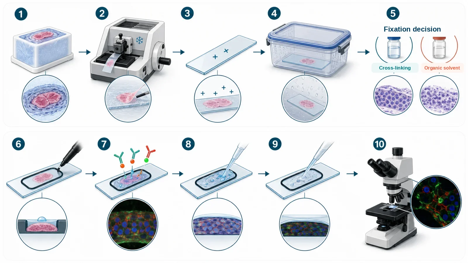
Before you start
| Check | Why it matters | Record |
|---|---|---|
| Tissue history | Collection delay, perfusion, pre-fixation, freezing rate, embedding, storage, and freeze-thaw exposure can change morphology, antigen accessibility, and background. | Collection time, fixation before freezing, cryoprotectant or OCT use, freezing method, block orientation, storage temperature, and freeze-thaw events. |
| Section thickness | Thickness affects mechanical stability, antibody penetration, optical sectioning, background, and apparent signal density. | Nominal thickness, cryostat temperature, blade condition, compression, chatter, folds, and tears. |
| Slide adhesion | Weak adhesion can cause partial or complete tissue loss during retrieval, incubation, or washing. | Slide type or coating, time before fixation, drying history, section lifting, and any tissue-dependent adhesion problem. |
| Fixation and permeabilization | Formaldehyde, alcohol, and acetone preserve different structures and expose different epitopes. | Antibody validation application, target compartment, structure that must be preserved, and candidate conditions to compare. |
| Baseline fluorescence | Intrinsic tissue fluorescence and fixation-related fluorescence can resemble specific signal and vary by channel. | Unstained section acquired in every planned channel using the same objective and comparable settings. |
| Acquisition plan | Changing exposure, gain, or processing between groups can create an apparent biological difference. | Microscope, objective, channels, exposure, gain, binning, z-step, flat-field method, and processing rules. |
Materials and reagents
| Category | Required | Selection notes |
|---|---|---|
| Tissue and embedding | Frozen tissue block, OCT or validated embedding medium, cryostat | Preserve orientation and keep the block at a stable cutting temperature appropriate for the tissue. |
| Slides | Positively charged or otherwise validated adhesive slides | Use one slide type consistently; highly fatty, mineralized, fragile, or previously fixed tissues may require additional optimization. |
| Wash buffer | PBS or TBS | Use one defined formulation across comparison groups. |
| Fixation route | Fresh 4% formaldehyde, ice-cold methanol, chilled acetone, or another validated route | Do not assume routes are interchangeable. Organic solvents can extract lipids and soluble material. |
| Permeabilization reagent | Validated detergent when intracellular access is required | Detergent may be included in blocking or antibody diluent, but stronger access can damage membranes and fine morphology. |
| Blocking and antibody diluent | Normal serum, BSA, or a validated commercial buffer | Normal serum is commonly matched to the host species of the secondary antibody. Avoid components incompatible with the detection chemistry. |
| Primary antibody | Antibody validated for frozen-tissue immunofluorescence or supported by appropriate local evidence | Record supplier, catalog number, clone, lot, concentration, dilution, and validation application. |
| Secondary antibody | Fluorophore-conjugated, host-specific secondary antibody | Use a secondary reactive to the primary-antibody host; highly cross-adsorbed reagents are preferred for multiplex work. |
| Mounting and handling | Hydrophobic barrier pen if compatible, humidified chamber, fine forceps, nuclear stain as needed, antifade mounting medium | Use enough volume to cover the tissue and prevent drying; confirm that barrier ink is compatible with the fluorophores and detection system. |
Starting conditions for frozen sections
These starting conditions are taken from current manufacturer protocols. Test them for the tissue, target, antibody, and preservation requirements of your experiment.
| Stage | Starting condition | Check |
|---|---|---|
| Fresh-frozen sectioning | Approximately 6–20 µm on a positively charged slide | A broad starting range used in a current CST frozen-tissue protocol. Select thickness from tissue mechanics, penetration, and imaging requirements. |
| Post-section formaldehyde fixation | Fresh 4% formaldehyde for approximately 15 minutes at room temperature | Common morphology-preserving route; usually requires a separate permeabilization decision for intracellular targets. |
| Optional cold methanol treatment | 100% methanol for approximately 10 minutes at −20°C or on ice | Fixes and permeabilizes but can extract lipids and alter membrane-rich or soluble structures. |
| Alternative acetone route | Chilled 100% acetone for approximately 10 minutes, only when supported by the antibody and tissue workflow | A commonly described frozen-section route, but morphology and target retention must be checked locally. |
| Blocking | Approximately 60 minutes at room temperature | Evaluate performance using the secondary-only control instead of assuming that longer blocking is always better. |
| Primary antibody | Manufacturer-recommended dilution, commonly overnight at 4°C | Titrate concentration and compare localization, positive control, and negative-control behavior. |
| Secondary antibody | Manufacturer-recommended dilution for approximately 1–2 hours at room temperature, protected from light | Match the primary host species and include a secondary-only section. |
| Washing | Three gentle washes of approximately 5 minutes between major labeling steps | Keep wash composition, duration, volume, and agitation consistent while avoiding direct force on the section. |
Procedure
- Document the tissue and block. Record collection delay, fixation before freezing, cryoprotection, embedding medium, orientation, freezing method, storage temperature, and freeze-thaw history.
- Equilibrate the block to the cryostat. Allow the block to reach a stable cutting temperature before trimming. Record compression, chatter, cracks, or curling before proceeding.
- Cut and mount representative sections. Place sections onto validated adhesive slides without stretching or trapping folds. Include adjacent sections for unstained, secondary-only, positive, and negative controls.
- Handle stored slides without condensation. If slides were stored frozen, keep them inside a closed container while they approach the required handling temperature. Open the container only after the external surface has equilibrated enough to reduce condensation on the tissue.
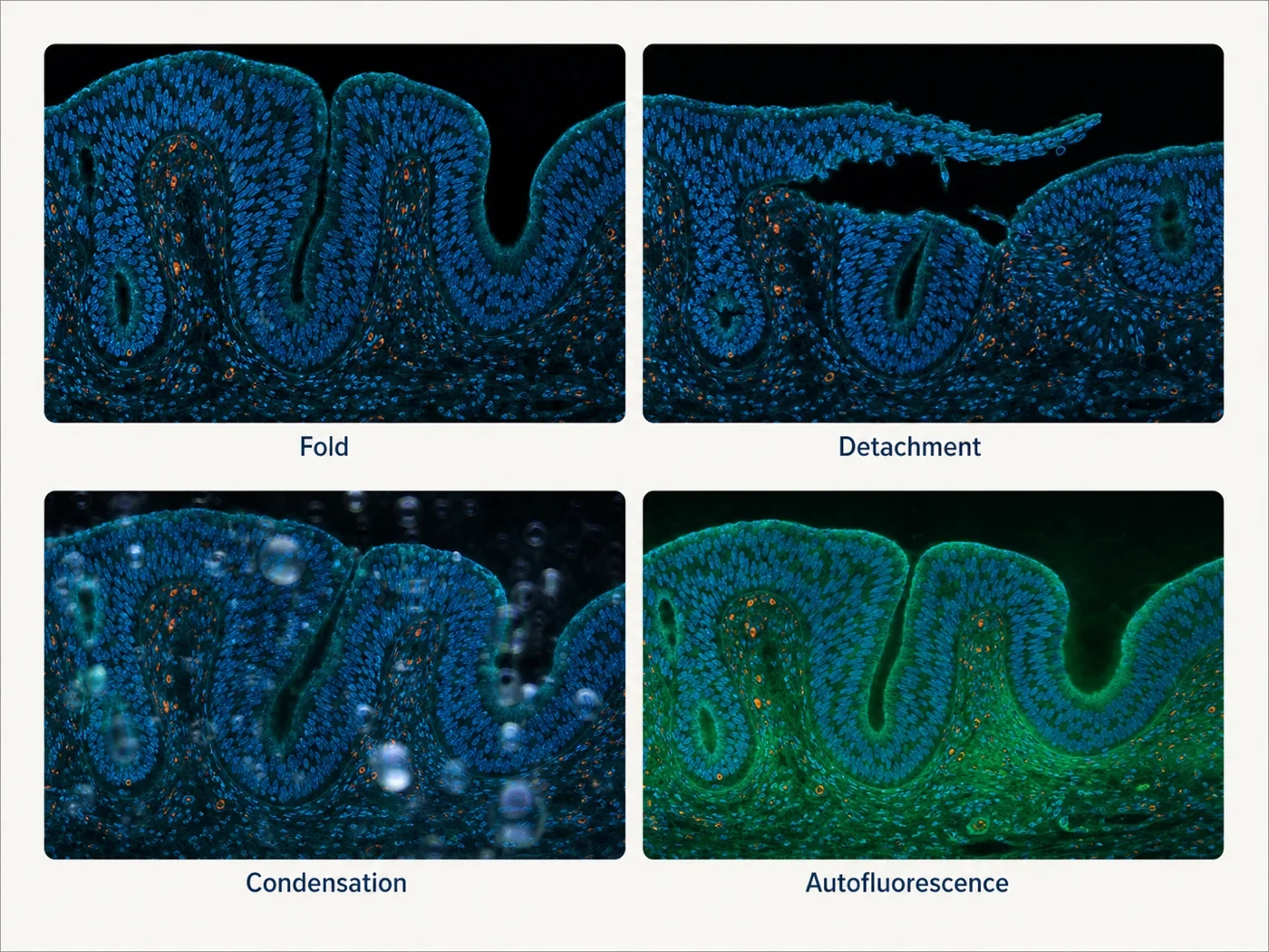
- Inspect adhesion before staining. Use brightfield inspection to identify folds, tears, incomplete transfer, compression, or lifting. Do not spend antibody on a section whose geometry or attachment already prevents interpretation.
- Fix the section. Use the preselected formaldehyde, methanol, acetone, or other validated condition. Keep reagent age, temperature, time, and volume consistent across comparison groups.
- Wash gently and keep the tissue covered. From the first aqueous wash onward, do not allow the section to dry. Avoid direct liquid jets and monitor edges for early lifting.
- Permeabilize when needed. Apply a validated detergent condition for intracellular targets when the fixation route has not already permeabilized the tissue. Use retrieval only when supported by the antibody and tissue preparation.
- Mark the staining area. Apply a compatible hydrophobic barrier after the section perimeter is dry enough for the pen, while keeping the tissue itself hydrated. Confirm that the reagent volume covers the complete section.
- Measure baseline fluorescence. Acquire the unstained section in every planned channel before interpreting antibody signal or selecting a quenching strategy.
- Block and incubate the primary antibody. Use a humidified chamber, prevent local evaporation, and record antibody identity, clone, lot, concentration, dilution, diluent, time, temperature, and volume.
- Wash consistently. Use gentle immersion or low-force exchange with the same number, duration, volume, and agitation for every comparable section.
- Incubate the fluorescent secondary antibody. Protect from light, match the primary-antibody host species, and control cross-reactivity when more than one primary host is used.
- Wash, counterstain, and mount. Remove unbound fluorophore, avoid bubbles and excessive coverslip pressure, and keep section geometry intact.
- Acquire controls first. Confirm section presence, baseline fluorescence, secondary background, known-positive signal, channel identity, and exposure below saturation before imaging experimental sections.
Minimum control set
| Control | What it checks | Limitation |
|---|---|---|
| Unstained section | How much tissue, fixative, pigment, embedding, or treatment-related fluorescence is present in each channel? | Antibody specificity or secondary-antibody background. |
| Secondary-only section | Does the fluorescent secondary antibody or detection chemistry create background without the primary antibody? | That the primary antibody recognizes only the intended target. |
| Known-positive tissue | Can the staining and acquisition workflow detect the expected tissue and subcellular pattern? | Target dependence in the experimental specimen. |
| Biological or genetic negative | Does signal decrease where the target is absent or substantially reduced? | All sources of tissue autofluorescence, bleed-through, or secondary background. |
| Adjacent or serial section | Is the pattern reproducible in neighboring anatomy or supported by an orthogonal stain? | Exact co-localization on the same section. |
| Single-color control | Does one fluorophore appear in another acquisition channel? | Target specificity or nonspecific antibody binding. |
Stop points and storage
- Frozen blocks: store under the validated conditions for the tissue and study. Record storage duration and every thaw or temperature excursion.
- Unfixed frozen slides: a validated laboratory workflow may allow storage at very low temperature, but humidity, condensation, and epitope stability must be controlled.
- After fixation and washing: a short pause in buffer at 4°C may be acceptable for many targets, but labile epitopes should be processed on a validated schedule.
- During primary incubation: overnight incubation at 4°C is a planned step. Maintain full coverage and humidity.
- After mounting: allow the mountant to cure as directed, then store flat at 4°C protected from light and record the interval before imaging.
- Avoid: repeated freeze-thaw cycles, opening a cold slide box in humid room air, allowing aqueous sections to dry, strong wash force, and storing comparison groups for different durations without validation.
Troubleshooting checkpoints
| Observation | Check first | Next guide |
|---|---|---|
| Partial or complete section loss | Charged-slide performance, section drying and storage history, tissue composition, retrieval severity, wash force, and edge lifting before staining. | Uneven-staining patterns |
| No or weak target signal | Known-positive tissue, section presence, acquisition channel and exposure, fixation route, access condition, and antibody validation application. | No-signal workflow |
| High diffuse background | Secondary-only section, exposure saturation, antibody titration, wash consistency, tissue drying, and hydrophobic-barrier compatibility. | High-background patterns |
| Strong channel-wide fluorescence | Unstained tissue in every channel, endogenous pigments, fixation history, treatment, and mounting medium. | Autofluorescence by channel |
| Patchy or edge-biased staining | Bubbles, incomplete coverage, local drying, folds, detachment, illumination gradient, and focus. | Uneven-staining patterns |
| Good signal but poor morphology | Freezing artifacts, cryostat temperature, blade condition, section compression, fixation route, detergent strength, and retrieval severity. | Fixation decision guide |
What to record
- Tissue identity, source, biological replicate, collection delay, and pre-freezing treatment
- Perfusion or immersion fixation before freezing, cryoprotection, embedding medium, orientation, and freezing method
- Block storage temperature, duration, thaw history, cryostat temperature, blade, and section thickness
- Slide type or coating, drying and storage history, and section-quality observations before staining
- Post-section fixation, permeabilization, or retrieval conditions with rationale
- Blocking, primary, and secondary antibody identifiers, lots, concentrations, dilutions, times, temperatures, and volumes
- Wash composition, count, duration, volume, agitation, and any section lifting
- Unstained, secondary-only, positive, negative, serial-section, and single-color controls as applicable
- Mounting medium, coverslip, curing and storage interval, microscope, objective, channels, exposure, gain, z-step, and processing
- Raw control images, uncropped representative fields, excluded sections, and reasons for exclusion
References and protocol sources
- Immunofluorescence protocol for frozen tissue (IF-F) Manufacturer protocol
Cell Signaling Technology
Charged slides, cryosection thickness, fixation, blocking, antibody incubation, washing, and mounting starting conditions.
- Tissue sample preparation guidance for immunostaining Manufacturer protocol
Thermo Fisher Scientific
OCT embedding, cryosection handling, coated slides, acetone fixation, and frozen-slide preparation.
- Overview of immunofluorescence techniques Technical guide
Cell Signaling Technology
Matching antibodies and protocols to biological material and sample processing.
- Frozen tissues with citrate antigen retrieval Manufacturer protocol
Cell Signaling Technology
Antigen retrieval as an optional antibody-specific branch for frozen sections.