Scope
This guide is for fixed-cell and tissue immunofluorescence in which label-independent fluorescence obscures, mimics, or reduces the dynamic range of a target signal. It covers cultured cells, frozen sections, FFPE sections, low-plex multiplex panels, conventional multichannel imaging, and spectral detection.
Autofluorescence is fluorescence generated by the specimen or its preparation without the intended fluorescent label. Distinguish it from nonspecific antibody binding, unbound fluorophore, bleed-through, detector noise, ambient light, and display processing before choosing a mitigation method.
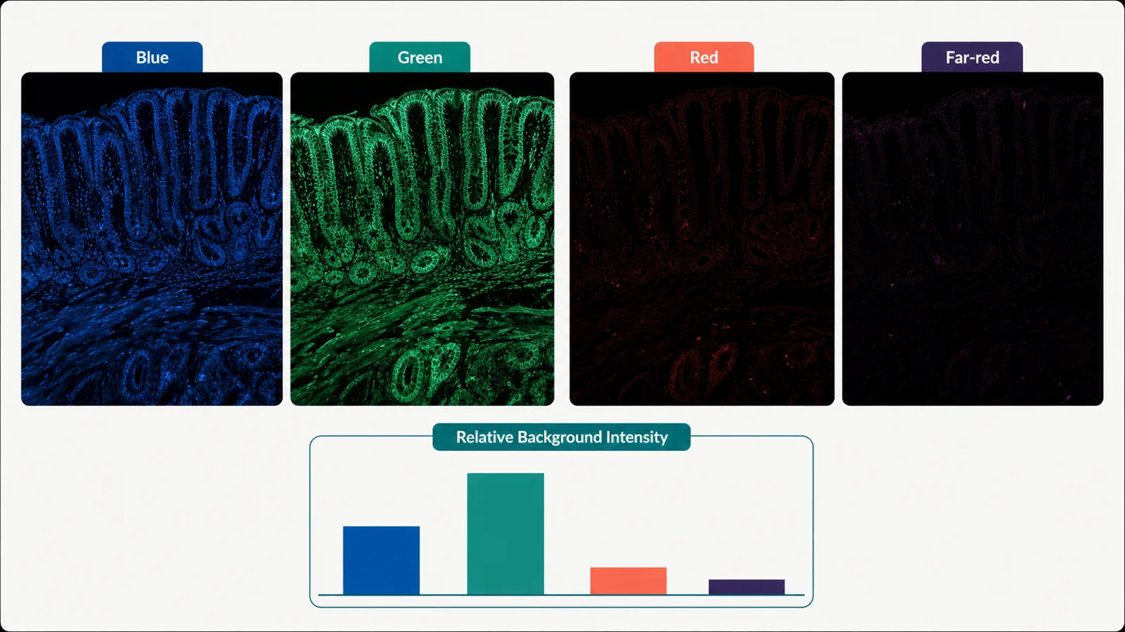
Quick check: is the fluorescence present without the label?
| Observation | Likely explanation | Check next |
|---|---|---|
| The same structure is bright in a preparation-matched unstained specimen | Specimen, fixation, processing, treatment, substrate, or mountant fluorescence is more likely than antibody binding | Acquire the unstained specimen through every final channel using the same objective and unsaturated settings |
| Unstained is dark, but the secondary-only or omission control is bright | The dominant source is the detection layer rather than autofluorescence | Return to secondary concentration, compatibility, Fc or immunoglobulin context, aggregates, and washing |
| Unstained is dark and only complete staining is bright | Primary-antibody concentration, specificity, broad true expression, or preparation-dependent off-target binding is more likely | Primary titration with known-positive and target-negative material |
| One single-color control appears in another channel | Cross-excitation, emission bleed-through, saturation, or spectral-unmixing error is more likely | Acquire that single-color specimen through every final channel |
| A granular or fibrous structure is bright across several excitation and emission combinations | A broad-spectrum endogenous or processing-associated source is plausible | Map the same structure in matched unstained material across channels or with a lambda scan |
| Background is present only after mounting, on plastic, or near adhesives and barriers | Material-associated fluorescence is more likely | Image blank substrate, coverslip, adhesive, barrier reagent, and mountant controls |
Map the autofluorescence before trying to remove it
- Prepare a matched unstained control. Match fixation, permeabilization, deparaffinization, retrieval, counterstain, mountant, storage, section age, and time to imaging.
- Use the final optical configuration. Record excitation, dichroic or beamsplitter, emission window, detector, objective, gain, exposure, z-step, and acquisition order.
- Acquire every planned channel below saturation. Do not use auto-exposure independently for each channel or sample.
- Map the spatial pattern. Record whether fluorescence follows lipofuscin-like granules, red blood cells, collagenous fibres, elastic structures, cell cytoplasm, tissue edges, necrosis, pigment, substrate, or mounting material.
- Measure representative regions. Include low- and high-background areas, not one selected field.
- Use spectral detection where available. Collect a matched autofluorescence reference with the same specimen preparation and instrument settings.
- Retain raw data. Preserve images before subtraction, denoising, unmixing, thresholding, or display adjustment.
A broad signal across several channels can suggest autofluorescence, but the key check is whether the same structure appears in matched unstained material.
Likely sources of autofluorescence
| Source class | Common pattern | Check | Limitation |
|---|---|---|---|
| Intracellular pigments and metabolites | Granular, vesicular, diffuse cytoplasmic, age- or tissue-dependent fluorescence across several channels | Matched unstained specimen, morphology, multiple channels, and tissue history | Appearance alone does not prove that the source is lipofuscin, flavin, NADH, or another molecule |
| Blood and heme-associated structures | Cell-shaped or vascular fluorescence that may appear in multiple channels | Unstained tissue, transmitted-light morphology, vascular or erythrocyte context, and serial sections | Do not interpret multichannel brightness near vessels as marker co-expression without controls |
| Extracellular matrix | Fibrous, vessel-wall, stromal, or connective-tissue pattern | Unstained section and structural correspondence with collagenous or elastic regions | Specific matrix component identity requires independent evidence |
| Aldehyde fixation | Diffuse or structure-associated fluorescence that increases with fixation chemistry, age, or exposure | Fresh, documented fixative and a controlled fixation comparison with matched unstained samples | Retrieval, washing, or blocking cannot necessarily reverse fixation-generated fluorophores |
| FFPE processing and retrieval | Channel-specific or diffuse tissue background after formalin fixation, paraffin processing, heat, or protease treatment | Unstained serial sections under no retrieval and the selected retrieval condition | Retrieval can change both target access and background at the same time |
| Drug, nanoparticle, reporter, pigment, or treatment | Background restricted to treated groups or specific intracellular structures | Treated and untreated unstained controls | Subtracting the mean background cannot resolve spatial overlap with the true label |
| Substrate, adhesive, barrier, coverslip, or mountant | Field-wide, edge-biased, surface, bubble-associated, or material-shaped fluorescence | Blank material controls and mounted unstained specimens | This is experimental-material background, not necessarily biological autofluorescence |
Check specimen-specific sources
Cultured cells
Compare cells without fluorescent labels, treated and untreated cells, blank vessel or coverslip, mounting medium, and fresh versus aged fixative. Check whether plastic, drug treatment, reporter expression, cell death, or overfixation creates the pattern.
Frozen sections
Check tissue age, blood content, intrinsic pigments, pre-fixation, post-section fixation, OCT or embedding material, section thickness, condensation, storage, mountant, and whether the same structures fluoresce in unstained serial sections.
FFPE sections
Check cold ischemia, formalin duration, processor history, block and section age, deparaffinization, retrieval, pigment, blood, collagen-rich areas, and formalin-associated background. Match every candidate mitigation to the actual FFPE preparation.
Multiplex panels
Acquire an autofluorescence reference and every single-color control through all final channels. Recheck the reference after changes to retrieval, fluorophores, detector windows, unmixing, cycle order, stripping, or amplification.
Reduce avoidable autofluorescence before adding a quencher
| Source | What to do | Keep in mind |
|---|---|---|
| Fixative | Use fresh, correctly prepared fixative and document concentration, age, temperature, duration, and delay to fixation | Do not weaken fixation so far that morphology or target retention fails |
| Specimen collection | Standardize blood content, handling delay, tissue thickness, treatment state, and storage where these variables affect background | Collection changes may alter biology and must be designed prospectively |
| Processing | Standardize deparaffinization, rehydration, retrieval, washing, section thickness, and drying prevention | More aggressive processing can create damage while reducing one background source |
| Materials | Use imaging-compatible substrates, coverslips, adhesives, barriers, and mountants verified in blank controls | A material that is acceptable in one channel may be bright in another |
| Panel design | Assign weak targets to channels with favorable measured signal-to-background on the actual microscope | Far-red is not automatically best; detector sensitivity and specimen background must support it |
| Acquisition | Use the minimum illumination and exposure needed, preserve unsaturated range, and avoid repeated pre-exposure | Lower exposure alone does not improve the underlying signal-to-background ratio |
Try optical or computational options before destructive treatment when possible
| Option | Potential benefit | Check |
|---|---|---|
| Channel reassignment | Moves a weak marker away from a high-background excitation or emission range | Matched singleplex and unstained comparison on the actual instrument |
| Narrower excitation or emission window | Rejects some background or neighbouring emission | Confirm adequate target signal and acceptable detector noise |
| Sequential acquisition | Reduces selected cross-excitation or simultaneous-channel contamination | Single-color controls; it does not remove autofluorescence or overlapping emission |
| Optical sectioning | Reduces out-of-focus haze from thick specimens | Same specimen thickness, z-position, objective, and raw data comparison |
| Spectral unmixing | Separates a measured autofluorescence component from fluorophore spectra | Matched autofluorescence and single-color references, unsaturated data, adequate spectral sampling, and residual inspection |
| Background modelling or subtraction | May improve visualization or some predefined quantitative workflows | Preserve raw data, apply the same rule to all groups, and demonstrate that the method does not erase target-positive structures |
Test quenching as another sample-preparation step
Chemical masking, chemical quenching, oxidation, and photobleaching can reduce selected autofluorescence sources, but their effects depend on tissue, fixation, wavelength, and treatment order. Test a quencher on the actual specimen rather than assuming a method from another tissue will transfer directly.
| Method | Potential target | Main risks | Comparison to run |
|---|---|---|---|
| Lipophilic dark dyes or commercial lipofuscin quenchers | Lipofuscin-like and lipid-associated broad fluorescence in selected tissues | Dark haze, masking of weak structures, reduction of true fluorophore signal, altered far-red background, precipitation, and sequence dependence | Serial sections: untreated unstained, treated unstained, untreated stained, and treated stained using the same acquisition |
| Copper-containing, ammonia/alcohol, trypan-blue, or other chemical treatments | Selected endogenous or fixation-associated sources | Incomplete quenching, shifted emission, tissue damage, reagent fluorescence, target loss, and incompatibility with later steps | Small tissue-specific screen with morphology and target-positive controls |
| Oxidative or light-based bleaching | Selected endogenous fluorophores before staining or in specialized iterative workflows | Epitope damage, fluorophore bleaching, oxidation, morphology change, long exposure, and uneven treatment | Apply only in a validated sequence and compare target detection, morphology, and spectral background before and after treatment |
| Commercial broad-spectrum quenching kits | Mixed or poorly defined tissue background | Unknown component specificity, lot effects, signal suppression, and platform incompatibility | Follow the current manufacturer protocol and validate on the actual specimen, fluorophores, retrieval, and imaging system |
Compare quenched and untreated samples
| Specimen | No quenching | Candidate quenching method | Keep constant |
|---|---|---|---|
| Unstained serial section | Baseline spectrum and morphology | Residual spectrum and morphology after treatment | Section thickness, preparation, mountant, storage, objective, filters, detector, and acquisition |
| Known-positive stained section | Target signal, background, localization, and dynamic range | Target retention, background reduction, localization, and morphology after treatment | Antibody clone and lot, concentration, fluorophore, incubation, washing, and acquisition |
| Target-negative stained section | Baseline target-independent signal | Residual target-independent signal after treatment | Detection method and processing |
| Single-color multiplex control | Baseline spillover and background | Effect of quencher on the fluorophore and neighbouring channels | Panel configuration and detector windows |
Required controls
| Control | What it checks | What failure looks like |
|---|---|---|
| Preparation-matched unstained specimen | What label-independent fluorescence exists in each channel? | The candidate target pattern is already present before labeling |
| Untreated and quencher-treated unstained serial sections | Does the intervention reduce the intended background without changing morphology? | Background shifts to another channel, remains patchy, or tissue darkens or deforms |
| Untreated and quencher-treated known-positive sections | Is true target signal, localization, and dynamic range preserved? | Target intensity or fine structure is lost along with background |
| Target-negative material | Does the remaining signal depend on the intended target? | The same pattern persists after quenching in negative material |
| Secondary-only or method-matched omission | Is the apparent background caused by the detection layer? | Background appears only after downstream detection reagents are added |
| One single-color control per fluorophore | Is another channel contaminated by the label rather than the specimen? | The structure appears in a channel lacking that fluorophore |
| Autofluorescence reference for spectral unmixing | Does the reference match the specimen and preparation used in the panel? | Residual halos, negative values, invented structures, or unstable separation |
| Morphology reference | Did prevention or quenching preserve the structure required for interpretation? | The slide becomes darker but architecture, membrane boundaries, nuclei, or fine processes are lost |
Choose one next test
| Current result | Next test | Keep unchanged |
|---|---|---|
| Unstained material is bright in one channel only | Compare one compatible alternate channel or narrower detection window | Keep antibody, specimen preparation, and biological sample fixed |
| Unstained material is bright across several channels | Map its spatial and spectral signature before selecting a tissue-supported quencher | Do not add amplification or raise antibody concentration |
| The same structure appears in a single-color control and a neighbouring channel | Reduce saturation, narrow the window, change acquisition grouping, or test an alternate fluorophore | Do not diagnose autofluorescence from the merged image |
| Background increases after formalin fixation or retrieval | Compare matched unstained serial sections using one milder validated preparation condition | Keep labeling and acquisition fixed |
| Lipofuscin-like granules obscure a weak target | Run a four-arm quencher comparison using untreated and treated unstained and stained serial sections | Do not adopt a published concentration from another tissue without validation |
| Spectral unmixing leaves halos or negative structures | Reacquire matched autofluorescence and single-color references below saturation | Do not tune the algorithm only on the final merged appearance |
When the autofluorescence problem is under control
- The label-independent spatial and spectral pattern is documented in a preparation-matched unstained specimen
- Bleed-through, detection-layer background, substrate fluorescence, and display artifacts have been separated from specimen autofluorescence
- The selected channel or mitigation leaves sufficient unsaturated dynamic range for the assigned target
- Known-positive material retains the expected target-dependent localization and signal after mitigation
- Target-negative material loses or substantially reduces the target pattern
- Single-color controls show acceptable contamination across all final channels
- Quenching or bleaching does not introduce new background, dark haze, unevenness, or loss of fine morphology
- Spectral unmixing uses matched references and produces acceptable residuals without invented structures
- The complete workflow is reproducible across the specimens, batches, devices, operators, and reagent lots required by the study
- Raw unstained, single-color, treated, untreated, and final images are retained with acquisition and processing metadata
Acceptable autofluorescence depends on what you need to measure. A background level adequate for locating a bright marker may still be unacceptable for a dim target, colocalization claim, rare-cell analysis, or quantitative intensity comparison.
Common autofluorescence patterns
| Pattern | Likely source | Check |
|---|---|---|
| Bright intracellular granules in several channels | Lipofuscin-like or other endogenous pigment | Matched unstained section, multiple channels, tissue age and cell-type context, and a validated quencher comparison |
| Vascular or blood-cell-shaped multichannel fluorescence | Red blood cells, heme-associated material, or pigment | Unstained serial section and transmitted-light or vascular morphology |
| Fibrous stromal fluorescence | Extracellular matrix such as collagenous or elastic structures | Unstained section and structural correspondence |
| Diffuse background stronger after aldehyde fixation | Fixation-associated fluorescence or altered tissue chemistry | Fresh fixative and one controlled fixation comparison with matched unstained specimens |
| Background stronger after retrieval | Retrieval-associated change, exposed endogenous structures, tissue damage, or increased collection of broad emission | No-retrieval and retrieved unstained serial sections under identical acquisition |
| Signal follows plastic, adhesive, mountant, or barrier geometry | Material fluorescence | Blank materials and mounted unstained controls |
| One fluorophore pattern appears in a neighbouring channel only after staining | Bleed-through or cross-excitation | Single-color control through every final channel |
| Quenched section develops dark haze or loses fine processes | Overmasking, precipitation, true-signal suppression, or tissue alteration | Untreated and treated known-positive serial sections with morphology reference |
Common autofluorescence mistakes
| Mistake | Why it fails | Better approach |
|---|---|---|
| “Anything visible in the unstained slide is lipofuscin.” | Blood, matrix, aldehyde products, treatment compounds, pigments, substrate, mountant, and other sources can look similar. | Describe the observed spatial and spectral pattern unless independent evidence identifies the source. |
| “Far-red always avoids autofluorescence.” | Detector sensitivity, excitation efficiency, tissue pigments, broad emitters, and fluorophore brightness vary. | Measure the unstained specimen and target signal on the actual instrument. |
| “A darker slide after quencher means the experiment improved.” | The treatment may also remove true fluorescence, mask fine structures, or create dark haze. | Compare untreated and treated unstained, positive, negative, and morphology controls. |
| “Sudan Black B, TrueBlack, or another quencher works at one universal concentration.” | Performance depends on tissue, fixation, wavelength, sequence, exposure, and target brightness. | Use the current product or published tissue-specific method as a starting point and validate a small matrix. |
| “Spectral unmixing removes the need for unstained controls.” | Unmixing requires a representative autofluorescence reference and valid single-color spectra. | Acquire matched references under the final configuration. |
| “Background subtraction proves a weak marker is real.” | Subtraction can create or erase structures and cannot establish target dependence. | Use positive, target-negative, omission, unstained, and single-color evidence. |
| “Photobleaching the background is harmless.” | Light and oxidation can alter epitopes, fluorophores, morphology, and different regions unequally. | Use only a validated sequence with treated and untreated controls. |
What to record
- Specimen identity, biological replicate, tissue or cell type, donor or age where relevant, treatment, region, blood or pigment context, and expected target pattern
- Fixation, fixative age, concentration, duration, temperature, delay, processing, section thickness, deparaffinization, retrieval, storage, and time to imaging
- Spatial description of label-independent fluorescence: granular, vascular, fibrous, diffuse, edge-biased, material-associated, or treatment-associated
- Unstained, secondary-only or omission, target-negative, known-positive, single-color, morphology, treated, untreated, and spectral-reference controls
- Microscope, objective, illumination, excitation, dichroic, emission windows, detector, exposure, gain, z-step, sequence, spectral sampling, saturation rule, and field-selection method
- Raw background intensity and representative regions in every planned channel before subtraction or unmixing
- Channel reassignment, filter or window changes, optical sectioning, unmixing, subtraction, quenching, bleaching, or other mitigation tested
- Quencher or bleaching reagent, supplier, catalog number, lot, preparation, sequence, concentration, duration, temperature, washes, and safety controls when used
- Effect on true target signal, target-negative material, morphology, fine structures, neighbouring channels, dynamic range, and residual background
- Raw-file location, processing parameters, residual inspection, rejected explanations, selected condition, repeat count, and revalidation triggers
References and protocol sources
- Background fluorescence and ways to reduce it Technical guide
Thermo Fisher Scientific
Separation of instrument, sample, vessel, medium, unbound-label, and treatment-related fluorescence; use of matched label-free controls and alternate channels.
-
Nawar Sakr, Olga Glazova, Liudmila Shevkova, Nikita Onyanov, Samira Kaziakhmedova, Alena Shilova, Maria V. Vorontsova, and Pavel Volchkov . International Journal of Molecular Sciences (2023) . DOI: 10.3390/ijms24043432
Broad, tissue-dependent autofluorescence from endogenous and fixation-related sources; spectral characterization; quencher comparisons; and the need to validate effects on specific fluorescence and tissue integrity.
-
Cell Signaling Technology
FFPE application validation, formalin-associated autofluorescence, sensitivity limits, and amplification considerations.
-
S. R. Yang, B. K. Maity, and S. Chong . The Journal of Physical Chemistry B (2023) . DOI: 10.1021/acs.jpcb.3c01658
Fixation-dependent redistribution, cross-linking and organic-solvent trade-offs, and interpretation limits.
- Selecting optical filters for fluorescence microscopy Technical guide
Thermo Fisher Scientific, Molecular Probes Handbook
Excitation filters, dichroic beamsplitters, emission filters, spectral separation, autofluorescence rejection, and instrument-aware optical trade-offs.
- Guide for using the Fluorescence SpectraViewer Technical guide
Thermo Fisher Scientific
Instrument-aware fluorophore compatibility and quantitative inspection of spectral overlap.
- Bleed-through in fluorescence imaging Technical guide
Thermo Fisher Scientific
Recognition of signal from one fluorophore in a neighboring channel and mitigation through compatible fluorophore and filter selection.
- The Society for Immunotherapy of Cancer statement on best practices for multiplex immunohistochemistry and immunofluorescence staining and validation Consensus guideline
Janis M. Taube and colleagues . Journal for ImmunoTherapy of Cancer (2020) . DOI: 10.1136/jitc-2019-000155
Singleplex-first panel development, marker-level controls, multiplex-to-singleplex concordance, and revalidation after panel changes.
- Society for Immunotherapy of Cancer: updates and best practices for multiplex immunohistochemistry and immunofluorescence image analysis and data sharing Consensus guideline
Janis M. Taube and colleagues . Journal for ImmunoTherapy of Cancer (2025) . DOI: 10.1136/jitc-2024-008875
Acquisition, segmentation, phenotyping, quality control, batch correction, and sharing of raw and processed outputs.