Scope
This guide is for fixed-cell and tissue immunofluorescence in which diffuse, structured, channel-wide, edge-biased, particulate, or target-independent fluorescence reduces interpretability. It covers cultured cells, frozen sections, FFPE sections, direct and indirect detection, and low-plex multiplex panels.
High background can begin before antibodies are added, come from the secondary or primary antibody, arise during sample preparation, or be introduced by the optical setup, acquisition, or display settings.
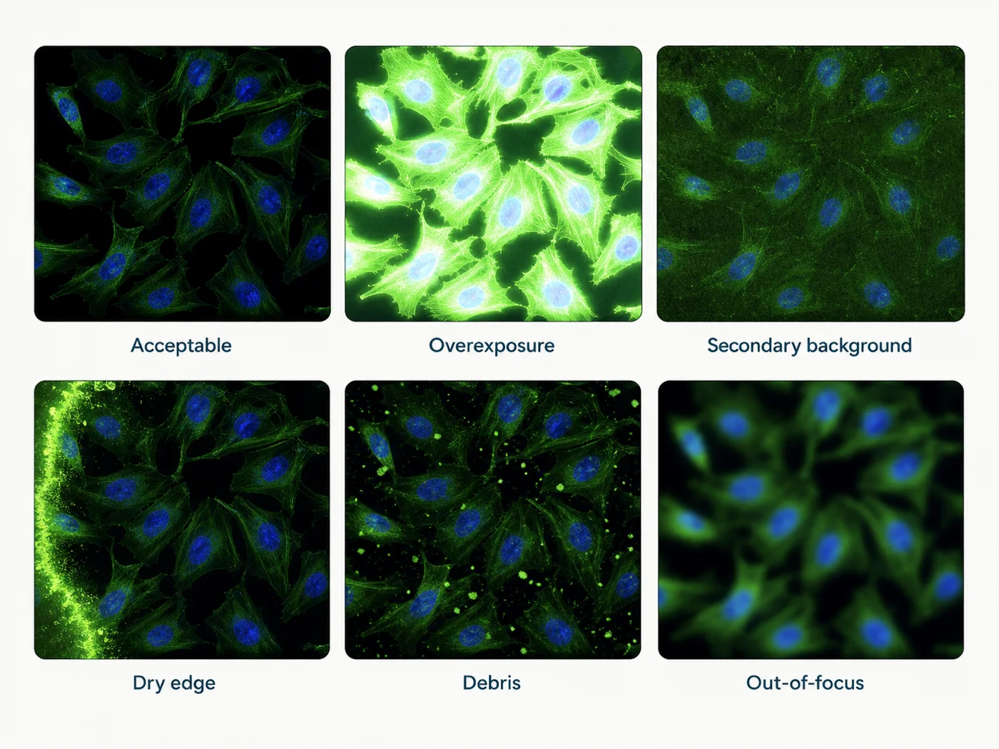
Quick check: when does the background first appear?
| Control result | Likely explanation | Check next |
|---|---|---|
| Unstained specimen is already bright in the affected channel | Intrinsic fluorescence, fixation or processing, treatment, substrate, mountant, or optical settings are more likely than antibody binding | Image a preparation-matched unstained specimen, blank substrate, and alternate channel below saturation |
| Unstained is acceptable but secondary-only control is bright | Secondary binding, endogenous immunoglobulin or Fc-related interactions, cross-reactivity, excess secondary, aggregation, or insufficient washing is more likely | Titrate the secondary, confirm species and cross-adsorption, inspect reagent quality, and compare a detection-method-matched control |
| Secondary-only is acceptable but complete staining is bright | Primary concentration, primary specificity, broad true expression, preparation-dependent off-target binding, or insufficient post-primary washing is more likely | Primary titration with known-positive and target-negative material while keeping detection and acquisition fixed |
| One single-color control appears in another channel | Cross-excitation, emission bleed-through, saturation, or spectral-unmixing error is more likely | Acquire that single-color specimen through every final channel using representative exposures |
| Only highly exposed or high-gain images look bright | Acquisition range, display scaling, detector noise, out-of-focus haze, or weak signal-to-background may be driving the appearance | Inspect raw values, exposure series, saturation indicators, optical sectioning, and the unstained control |
| Background is confined to edges, bubbles, debris, or isolated regions | Drying, coverage, precipitate, detachment, folds, contamination, or illumination nonuniformity is more likely than uniform antibody failure | Map the physical pattern before changing antibody concentration or blocking |
Look at the background pattern
| Pattern | Likely causes | Check |
|---|---|---|
| Uniform field-wide haze | High exposure, concentrated antibody, inadequate washing, specimen autofluorescence, out-of-focus light, or fluorescent substrate | Compare unstained, secondary-only, and exposure series using the same field and objective |
| Bright tissue or cell type in every channel | Intrinsic fluorescence, pigment, blood products, lipofuscin, collagen or elastin, fixation-related fluorescence, or spectral contamination | Unstained serial section through every channel and single-color controls |
| Diffuse cytoplasmic or nuclear haze only with primary antibody | Excess primary, off-target binding, damaged membranes, over-permeabilization, over-retrieval, or target-independent nuclear binding | Primary titration, target-negative material, morphology, and one milder preparation condition |
| Bright edges, rings, or hydrophobic-barrier boundary | Local drying, meniscus concentration, insufficient volume, evaporation, or incomplete coverage | Review incubation volume, chamber humidity, barrier geometry, and whether the specimen remained wet |
| Discrete bright particles | Antibody or fluorophore aggregates, precipitated blocking reagent, contaminated buffer, dust, tissue debris, or mounting artifacts | Inspect reagents and slide before staining, compare filtered or freshly prepared reagent where appropriate, and check whether particles are present in controls |
| Patchy regions following folds or thickness | Uneven reagent access, tissue thickness, incomplete deparaffinization, folds, section lifting, focus, or illumination gradient | Transmitted-light morphology, z-position, section quality, and an unstained serial section |
| Background rises from first to last field | Display auto-scaling, detector drift, focus changes, drying during acquisition, or inconsistent illumination | Fixed acquisition settings, field-order reversal, and a stable fluorescent reference |
Step 1: measure background before antibodies
Acquire a preparation-matched unstained specimen in every planned channel using the final objective, filters or detector windows, and a documented unsaturated acquisition rule.
- Match fixation, permeabilization, deparaffinization, retrieval, counterstain, mountant, storage, and time to imaging
- Include treated and untreated unstained material when drugs, reporters, nanoparticles, pigments, or diet may fluoresce
- Inspect representative tissue regions rather than one visually favorable field
- Image blank substrate, coverslip, adhesive, barrier reagent, or mounting medium when the background follows materials rather than biology
- Record channel-specific raw intensity and spatial distribution before any background subtraction
Step 2: check the detection reagents
| Detection method | Background control | What a positive control suggests |
|---|---|---|
| Indirect IF | Secondary-only or primary-omission control using the same secondary concentration and incubation | Secondary binding, Fc-related interactions, endogenous immunoglobulin, cross-reactivity, aggregates, or excessive secondary exposure |
| Directly conjugated primary | Method-matched omission control and target-negative material | Conjugate background, nonspecific primary binding, free dye or aggregates, or specimen autofluorescence |
| Biotin or streptavidin detection | Detection reagent without the intended target-binding layer and an endogenous-biotin control where relevant | Endogenous biotin or nonspecific avidin or streptavidin interactions |
| Amplified detection | Each omitted amplification layer in the validated sequence | Amplifier deposition, overdevelopment, endogenous activity, or sequence error |
| Same-host or sequential multiplex | Order-specific omission controls after each blocking, stripping, or Fab step | Residual binding sites, cross-detection of an earlier primary, incomplete stripping, or cumulative background |
Secondary-antibody checks
- Confirm host species, immunoglobulin class, fragment, conjugate, fluorophore, and intended primary compatibility
- Confirm cross-adsorption claims against the actual species present in the panel and specimen
- Titrate the secondary independently while keeping the primary and acquisition unchanged
- Inspect for precipitation, repeated freeze-thaw, light damage, contamination, or incorrect storage
- Use the same incubation volume, humidity, wash sequence, and exposure as the complete assay
Step 3: check whether the background comes from imaging
| Optical factor | How it raises apparent background | Check |
|---|---|---|
| Exposure, gain, or detector voltage | Raises specimen background, detector noise, and weak off-target signal together | Unsaturated exposure series with the same specimen and controls |
| Wide emission window | Collects more intended signal but also more autofluorescence and neighboring emission | Compare a narrower compatible window or filter using single-color and unstained controls |
| Cross-excitation and bleed-through | Bright fluorophore contributes to another channel | Acquire every single-color specimen through every final channel |
| Out-of-focus fluorescence | Widefield or thick specimens contribute haze above and below the focal plane | Review section thickness, z-position, optical sectioning, deconvolution inputs, and a thinner specimen where justified |
| Illumination nonuniformity | Field gradients can resemble uneven biological staining | Reference slide or flat-field assessment and specimen rotation |
| Display scaling or processing | Auto-contrast, local contrast, background subtraction, denoising, or clipping can exaggerate or hide background | Inspect raw channels with a documented common display rule |
Sequential acquisition can reduce some channel interaction, but it does not remove overlapping emission, autofluorescence, saturation, detector noise, or invalid single-color references.
Step 4: titrate the primary antibody
Titrate antibody concentration against both known-positive and target-negative material. Look for the best target-dependent signal relative to matched background, not the highest absolute intensity.
| Observation | Comparison to try | What this result means |
|---|---|---|
| Background falls as primary concentration is reduced while the expected pattern remains | Primary dilution series with fixed secondary and acquisition | The original primary concentration was unnecessarily high |
| Expected and off-target patterns fall together | Target-negative material and an independent antibody or genetic control | Concentration alone may not solve a specificity problem |
| Background is unchanged across primary concentrations | Secondary-only, unstained, and exposure controls | The dominant source may not be the primary antibody |
| Background appears only after a lot or conjugate change | Old and new lots or unconjugated and conjugated equivalents on the same specimen | Lot, conjugation, aggregation, or storage may have changed performance |
| Signal follows an unexpected compartment in positive and negative material | Application-specific validation, independent antibody, and preparation comparison | Do not label the pattern as biological localization from concentration optimization alone |
Western blot bands, expected localization, isotype controls, or peptide absorption may provide limited information but do not replace target-dependent immunofluorescence validation in the actual preparation.
Step 5: check blocking, diluent, and washing
| Variable | How it can affect background | Comparison to try |
|---|---|---|
| Blocking reagent | Insufficient surface occupancy, incompatible serum species, Fc-related interactions, or added endogenous immunoglobulin can alter background | Use the antibody-validated blocker and one justified alternative while keeping antibodies and acquisition fixed |
| Antibody diluent | Protein concentration, detergent, salt, pH, preservatives, and carrier components can change binding and aggregation | Compare the validated diluent with one documented alternative |
| Detergent during incubation | May reduce some nonspecific interactions but can also increase access, extraction, or cumulative specimen damage | Record total detergent exposure and compare one concentration or duration at a time |
| Wash composition | Insufficient washing leaves unbound reagent; harsh washing can damage or detach the specimen | Change wash duration, count, volume, or agitation one variable at a time |
| Incubation volume and humidity | Evaporation concentrates antibody and produces edge artifacts | Use complete coverage, controlled humidity, and documented chamber conditions |
| Reagent quality | Microbial contamination, precipitate, expired protein, or incorrect pH can create particulate or diffuse background | Inspect and prepare fresh documented reagent where degradation is plausible |
Step 6: check sample-preparation causes
| Preparation step | How it can raise background | Comparison to try |
|---|---|---|
| Fixation | Aldehyde-associated fluorescence, overfixation, delayed fixation, redistribution, or altered epitope access | Preparation-matched unstained control and one antibody-supported fixation comparison |
| Permeabilization | Excessive membrane disruption exposes new binding sites, extracts structures, and increases diffuse access | Compare no detergent with one milder condition while keeping fixation and antibodies fixed |
| FFPE deparaffinization | Residual paraffin causes uneven wetting, patchy fluorescence, and poor reagent exchange | Repeat a documented clearing and rehydration sequence before changing antibodies |
| Antigen retrieval | Excess heat or protease can increase autofluorescence, expose target-independent sites, damage tissue, and lift sections | Compare no retrieval or a milder validated condition with the current route |
| Section or specimen thickness | More fluorescent material and out-of-focus signal increase apparent background | Compare a validated thinner section or optical sectioning strategy |
| Mounting and storage | Autofluorescent mountant, poor refractive matching, oxidation, light exposure, or seal failure changes background and signal | Image a freshly mounted matched control and standardize storage and time to imaging |
Check multiplex-specific causes
- Return each marker to singleplex. Confirm that the background is absent or acceptable before panel assembly.
- Acquire every single-color control through every final channel. Match labeling density and acquisition to the complete panel.
- Check order and detection method. Same-host primaries, residual secondary-binding sites, stripping, amplification, and cumulative retrieval can add background.
- Add one marker or layer at a time. Identify the first panel addition that changes background or localization.
- Inspect raw channels before the merge. A composite can hide which fluorophore, marker, or cycle created the problem.
- Recheck the panel after changes. Fluorophore reassignment, antibody lot, sequence, retrieval, stripping, detector window, or analysis changes can alter background.
Choose one next test
| Current result | Next test | Keep unchanged |
|---|---|---|
| Unstained specimen is bright | Compare one alternative compatible channel or preparation-matched fixation or retrieval condition | Do not change antibody concentrations first |
| Secondary-only control is bright | Run a secondary dilution series and confirm species, cross-adsorption, reagent quality, and Fc or immunoglobulin context | Keep the primary omitted and acquisition fixed |
| Only complete staining is bright | Titrate the primary against known-positive and target-negative material | Keep secondary concentration, preparation, and acquisition fixed |
| One single-color control contaminates another channel | Reduce saturation, narrow the window, change sequential grouping, or test an alternate compatible fluorophore | Do not diagnose antibody specificity from the contaminated merge |
| Background follows stronger permeabilization or retrieval | Compare one milder access or retrieval condition | Keep antibody concentrations and imaging unchanged |
| Background is edge-biased or particulate | Correct volume, humidity, drying, bubbles, precipitate, or reagent cleanliness and repeat the same staining | Do not redesign the entire protocol |
When the background problem is under control
- Unstained background leaves sufficient usable dynamic range in every required channel
- Detection-layer controls remain below the predefined background threshold
- Known-positive material retains the expected target-dependent spatial pattern below saturation
- Target-negative material loses or substantially reduces the pattern without a new preparation-dependent signal
- Single-color controls show acceptable contamination across all other channels
- Specimen morphology, membrane boundaries, organelles, nuclei, tissue architecture, and section adhesion required by the question remain interpretable
- The selected antibody concentrations, blocking, washing, preparation, and acquisition are documented and reproducible
- The result remains acceptable across the specimens, days, operators, reagent lots, and instruments required by the study
- Raw channels and controls are retained without relying on undisclosed background subtraction or selective display scaling
Acceptable background depends on what you need to measure. A condition that works for a bright cell-type marker may still be inadequate for a dim or co-expressed target.
Common high-background patterns
| Pattern | Likely cause | Check |
|---|---|---|
| All channels are bright in unstained tissue | Intrinsic fluorescence, fixation, pigment, mountant, substrate, or exposure | Preparation-matched unstained control and alternate channel or optical configuration |
| Only antibody channels are bright, including secondary-only | Detection-layer concentration, compatibility, Fc or endogenous immunoglobulin interactions, or aggregation | Secondary titration and detection-method check |
| One target channel is diffusely bright while other antibody channels are clean | Primary-specific concentration, off-target binding, fluorophore aggregate, or channel-specific background | Primary titration, target-negative material, direct versus indirect comparison, and single-color control |
| Bright rings at cell or tissue edges | Drying, meniscus concentration, membrane damage, or edge-focused illumination | Humidity, volume, coverage, morphology, and unstained control |
| Bright dots outside cells or tissue | Aggregate, precipitate, dust, debris, or mounting artifact | Inspect reagent and blank slide; determine whether dots appear in omission controls |
| Background increases after antigen retrieval | Autofluorescence, tissue damage, exposed nonspecific sites, or excessive retrieval | Unstained and secondary-only serial sections with no retrieval and a milder condition |
| Background appears only after multiplex assembly | Spectral contamination, same-host conflict, cumulative treatment, incomplete stripping, or amplification carryover | Singleplex references, single-color controls, order-specific omissions, and incremental panel reconstruction |
Common troubleshooting mistakes
| Mistake | Why it fails | Better approach |
|---|---|---|
| “High background means blocking was insufficient.” | Autofluorescence, bleed-through, saturation, aggregates, drying, preparation damage, and primary off-target binding can look similar. | Use unstained and detection-layer controls to locate the first abnormal step. |
| “Increase blocking time and wash time together.” | The effective change cannot be identified, and harsh washing may damage the specimen. | Change one blocker or wash variable at a time. |
| “Reduce exposure until the background disappears.” | True target signal may disappear at the same time, while the underlying signal-to-background remains poor. | Compare raw target-dependent signal and matched background across an unsaturated exposure range. |
| “Secondary-only is clean, so the antibody is specific.” | It tests the downstream detection layer, not target dependence of the primary antibody. | Add target-negative, genetic, independent-antibody, or other application-specific evidence. |
| “Far-red always solves tissue autofluorescence.” | Detector sensitivity, fluorophore excitation, tissue pigments, and actual background vary. | Measure unstained background and target signal on the real instrument. |
| “Sequential scanning eliminates background.” | It cannot remove intrinsic fluorescence, overlapping emission, saturation, nonspecific binding, or invalid references. | Use compatible fluorophores and complete controls first. |
| “Background subtraction can rescue the experiment.” | Post-processing cannot restore lost specificity, dynamic range, morphology, or saturated pixels. | Resolve the experimental source and preserve raw data. |
What to record
- Specimen identity, biological replicate, cell line or tissue, treatment, region, preparation, thickness, and expected target pattern
- Spatial pattern of background: uniform, compartmental, edge-biased, particulate, patchy, channel-specific, or multiplex-dependent
- Unstained, secondary-only or omission, target-negative, known-positive, single-color, singleplex, morphology, and order-specific controls
- Primary and detection reagents with supplier, catalog number, clone, lot, host, isotype, conjugate, fluorophore, concentration, dilution, storage, and preparation
- Blocking reagent, serum species, protein concentration, antibody diluent, detergent, salt, pH, incubation volume, humidity, time, and temperature
- Wash buffer, count, duration, volume, agitation, specimen damage, drying, bubbles, precipitate, and contamination observations
- Fixation, permeabilization, deparaffinization, rehydration, retrieval, mounting, storage, and time to imaging
- Microscope, objective, illumination, excitation, dichroic, emission window, detector, exposure, gain, binning, z-step, order, optical sectioning, and saturation rule
- Raw-file location, background and signal measurement regions, processing, display range, rejected explanations, and the next test selected
- Variables changed in each repeat, acceptance threshold, final conclusion, repeat count, and revalidation trigger
References and protocol sources
- Immunofluorescent staining of intracellular antigens on cultured cells Manufacturer protocol
Thermo Fisher Scientific
Cultured-cell fixation, washing, blocking, antibody incubation, mounting, and storage starting conditions.
- Immunofluorescence protocol for cell-based imaging Manufacturer protocol
Cell Signaling Technology
Product-specific validation and formaldehyde-versus-methanol sample preparation.
- Immunofluorescence protocol for frozen tissue (IF-F) Manufacturer protocol
Cell Signaling Technology
Charged slides, cryosection thickness, fixation, blocking, antibody incubation, washing, and mounting starting conditions.
-
Cell Signaling Technology
FFPE application validation, formalin-associated autofluorescence, sensitivity limits, and amplification considerations.
-
S. R. Yang, B. K. Maity, and S. Chong . The Journal of Physical Chemistry B (2023) . DOI: 10.1021/acs.jpcb.3c01658
Fixation-dependent redistribution, cross-linking and organic-solvent trade-offs, and interpretation limits.
-
Richard W. Burry . Journal of Histochemistry & Cytochemistry (2011) . DOI: 10.1369/jhc.2010.956920
Functional separation of primary-antibody, secondary-antibody, and label controls; interpretation limits of omission and absorption controls.
- Controls for immunohistochemistry: The Histochemical Society’s standards of practice for validation of immunohistochemical assays Consensus guideline
Stephen M. Hewitt, Denis G. Baskin, Charles W. Frevert, William L. Stahl, and Eduardo Rosa-Molinar . Journal of Histochemistry & Cytochemistry (2014) . DOI: 10.1369/0022155414545224
Positive and negative control requirements, limits of primary-omission and absorption controls, and application-specific evidence for antibody specificity.
- Selecting optical filters for fluorescence microscopy Technical guide
Thermo Fisher Scientific, Molecular Probes Handbook
Excitation filters, dichroic beamsplitters, emission filters, spectral separation, autofluorescence rejection, and instrument-aware optical trade-offs.
- Bleed-through in fluorescence imaging Technical guide
Thermo Fisher Scientific
Recognition of signal from one fluorophore in a neighboring channel and mitigation through compatible fluorophore and filter selection.
- Guide for using the Fluorescence SpectraViewer Technical guide
Thermo Fisher Scientific
Instrument-aware fluorophore compatibility and quantitative inspection of spectral overlap.
- Multiplexed IHC staining with primary antibodies conjugated to fluorophores Manufacturer protocol
Thermo Fisher Scientific
Directly conjugated primary-antibody workflow, single-color controls, negative controls, coverage, and antibody titration.
- The Society for Immunotherapy of Cancer statement on best practices for multiplex immunohistochemistry and immunofluorescence staining and validation Consensus guideline
Janis M. Taube and colleagues . Journal for ImmunoTherapy of Cancer (2020) . DOI: 10.1136/jitc-2019-000155
Singleplex-first panel development, marker-level controls, multiplex-to-singleplex concordance, and revalidation after panel changes.