Scope
This guide covers how to choose and compare fixation methods for adherent cultured cells and cryosections used in immunofluorescence. It also explains how previous fixation affects frozen and FFPE tissue.
Fixation, permeabilization, antigen retrieval, and staining are related but distinct steps. A detergent cannot reverse fixation damage, and antigen retrieval is not a substitute for documenting how a specimen was fixed.
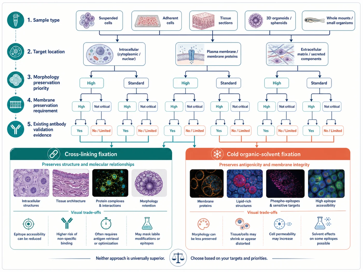
How fixation affects staining
There is no universally best fixative. Choose a condition that preserves the structure you need to see, keeps the expected target localization, allows antibody access, and gives acceptable background with the appropriate controls.
Target retention
Soluble, weakly associated, phase-separated, and rapidly moving proteins may redistribute or be lost before fixation is complete.
Structural preservation
Membranes, lipids, cytoskeleton, organelles, nuclei, and tissue architecture respond differently to cross-linking and solvent precipitation.
Epitope accessibility
Cross-linking can stabilize structure while restricting antibody access or changing the conformation of some epitopes.
Fluorescence background
Aldehyde fixation can increase autofluorescence, especially when fixation is strong or prolonged and in intrinsically fluorescent tissue.
Compare fixation methods
| Method | Potential strengths | Main risks | When to use |
|---|---|---|---|
| Aldehyde cross-linking, commonly fresh 4% formaldehyde | Often preserves overall morphology, membrane-rich structures, and spatial relationships better than organic solvents. | Can mask or restrict access to some epitopes, increase autofluorescence, and still permit redistribution before cross-linking is complete. Intracellular targets usually require a separate permeabilization decision. | The antibody and specimen support this preparation, and morphology or membrane retention is important. |
| Cold methanol | Rapidly precipitates proteins and simultaneously permeabilizes. It can work well for some cytoskeletal proteins and epitopes that perform poorly after aldehyde fixation. | Extracts lipids, can alter membranes and organelles, may shrink or distort cells, and can remove or redistribute soluble material. | Application-specific evidence or a controlled comparison shows acceptable target pattern and morphology. |
| Cold acetone | Rapid precipitation and permeabilization; used in some frozen-section workflows. | Strong extraction of lipids and soluble components, tissue brittleness, shrinkage, and morphology loss. | The antibody and specimen workflow specifically support it and tissue retention is verified. |
| Mixed or sequential fixation | May balance rapid immobilization, epitope access, and structural preservation for a specialized target. | Adds variables and can combine the disadvantages of both routes. Order and timing become part of the assay. | A published or product-specific method is being reproduced, or a deliberate comparison demonstrates a clear benefit. |
| Glutaraldehyde-containing fixation | Strong structural stabilization for specialized applications. | High autofluorescence and restricted antigen access can make routine immunofluorescence difficult. | The target or imaging method requires it and a validated quenching and staining workflow is available. |
Consider the specimen type
| Specimen | Key fixation question | Record before staining |
|---|---|---|
| Adherent cultured cells | Can the target and required structure be preserved before washing, detachment, or redistribution? | Live-cell morphology, confluence, treatment timing, expected target mobility, substrate, and antibody validation route. |
| Fresh-frozen cryosection | Was tissue fixed before freezing, and is post-section fixation needed? | Collection delay, perfusion or immersion fixation, cryoprotection, freezing method, section thickness, slide adhesion, and storage history. |
| Previously fixed cryosection | Will additional fixation improve retention or simply increase masking and background? | Original fixative, concentration, duration, tissue dimensions, delay to fixation, and prior storage. |
| FFPE tissue | How did pre-analytical formalin fixation and processing affect antigen accessibility and fluorescence? | Cold ischemia, fixative, fixation duration, processor program, block age, section age, and known-positive FFPE material. |
For FFPE specimens, changing retrieval cannot recreate an unfixed or differently fixed sample. Record the fixation history and optimize retrieval separately for the target.
How to choose a fixation method
- Identify the structure that must remain intact. State whether the conclusion depends on a membrane boundary, lipid droplet, soluble pool, filament, organelle, nuclear compartment, tissue architecture, or relative abundance.
- Check antibody validation in the same application. Validation in western blotting, FFPE IHC, frozen tissue, or cultured-cell IF is not automatically transferable to another preparation.
- Consider extraction and redistribution. Check whether the target is soluble, membrane-associated, lipid-dependent, weakly bound, phase-separated, or rapidly transported.
- Keep fixation and permeabilization separate when testing conditions. Decide whether the fixation method itself provides access or whether a later detergent step is needed. Do not increase detergent while simultaneously changing fixation.
- Measure baseline fluorescence. Acquire an unstained specimen in every planned channel before accepting a fixation condition or applying a quenching method.
- If the best condition is unclear, compare two reasonable methods first. Avoid a large matrix in which fixation, permeabilization, antibody concentration, and acquisition all change together.
Compare two fixation methods
For cultured cells, a useful first comparison can include one aldehyde method and one solvent method when both are supported by the antibody or target biology. These are starting conditions, not universal optima.
| Variable | Condition A | Condition B | Keep constant |
|---|---|---|---|
| Fixation | Fresh 4% formaldehyde, commonly 10–15 minutes at room temperature | Ice-cold 100% methanol, commonly 5–15 minutes at −20°C or on ice | Cell state, treatment, substrate, wash method, reagent volume, and time from medium removal to fixation |
| Access | One validated detergent condition after fixation when intracellular access is required | No separate detergent unless the application specifically requires it | Do not compare multiple detergent strengths in the first fixation experiment |
| Labeling | Same primary clone and lot, antibody concentration, secondary antibody, incubation, wash, counterstain, and mounting medium | Prepare dilutions together and stain in parallel | |
| Acquisition | Same objective, optical configuration, exposure logic, gain, z-step, and processing | Acquire below saturation and retain raw channels | |
Required controls
- Known-positive material processed under each fixation condition
- Target-negative biological or genetic material where feasible
- Unstained specimen under each fixation condition and every acquisition channel
- Primary-omission or secondary-only control for indirect detection
- Brightfield or transmitted-light morphology before fixation and after staining when applicable
- At least one independent repeat before selecting a condition for quantitative work
Before choosing a fixation condition
Choose the condition that preserves the expected localization and morphology while keeping background acceptable for the experiment.
- Expected cellular or tissue morphology remains interpretable
- Membranes, lipid-dependent structures, organelles, filaments, or nuclei required by the question remain intact
- Known-positive material shows the expected spatial pattern below detector saturation
- Target-negative material shows the expected loss or reduction without a new fixation-dependent pattern
- Unstained and detection-layer controls define acceptable background in every relevant channel
- The target pattern is not explained by extraction, collapse, edge staining, precipitation, or redistribution
- The result is reproducible across the required specimens, days, operators, and reagent lots
- The selected condition remains compatible with downstream permeabilization, multiplexing, mounting, and quantitative analysis
Recognize fixation-related artifacts
| Observation | Possible fixation explanation | Check |
|---|---|---|
| Expected morphology but weak or absent target signal | Epitope masking, excessive fixation, or insufficient post-fixation access | Known-positive performance, shorter aldehyde exposure, an antibody-supported solvent route, and a separate permeabilization comparison |
| Diffuse signal or loss of a soluble compartment | Redistribution before immobilization, extraction during washing, or underfixation | Reduce delay to fixation, compare faster immobilization, inspect live-cell localization where feasible, and evaluate the negative control |
| Membrane, lipid droplet, or organelle collapse after methanol or acetone | Solvent extraction of lipids and associated structural changes | Compare an aldehyde route while keeping antibody and acquisition constant |
| Bright background in unstained material | Intrinsic fluorescence or aldehyde-associated autofluorescence | Acquire unstained controls by channel, compare fixation duration, and determine whether the pattern predates antibody staining |
| Cell shrinkage, gaps, or distorted boundaries | Solvent precipitation, osmotic stress, drying, or harsh fluid exchange | Review transmitted-light images, reagent temperature, exchange method, and whether the artifact appears before antibody incubation |
| Good signal but different localization between fixatives | One condition may extract, redistribute, mask, or expose a different pool | Do not average the results. Compare target-negative material, independent antibodies, live-cell evidence, and expected compartment biology |
Common fixation mistakes
| Mistake | Why it fails | Better approach |
|---|---|---|
| Using the same fixative for every antibody | Different epitopes and target pools respond differently to cross-linking, precipitation, and extraction. | Start from application-specific validation and compare plausible routes when the evidence is incomplete. |
| Calling methanol a membrane-preserving fixative | Methanol extracts lipids and can distort membrane-dependent structures. | Describe it as a precipitating fixative that also permeabilizes, with target- and structure-dependent performance. |
| Changing fixation, detergent, and antibody concentration together | The cause of any improvement or failure becomes unknowable. | Compare fixation first with downstream variables held constant, then optimize access separately. |
| Selecting the brightest image | Brightness can come from background, exposed off-target epitopes, saturation, or morphology loss. | Use positive, negative, unstained, and detection controls plus morphology and reproducibility. |
| Assuming retrieval reverses overfixation | Retrieval may improve access to some epitopes but cannot restore extracted, redistributed, or destroyed structures. | Document fixation history and optimize retrieval as a separate variable. |
| Quenching autofluorescence without measuring it first | A quenching treatment can alter specific signal, color balance, or morphology and may hide the source of background. | Measure unstained material by channel before and after any proposed treatment. |
What to record for fixation
- Specimen identity, source, cell state or tissue history, treatment, biological replicate, and time to fixation
- Fixative chemical identity, supplier, stock, preparation, buffer, pH where relevant, concentration, age, and temperature
- Fixation start time, duration, reagent volume, mixing or immersion method, specimen dimensions, and wash sequence
- Pre-fixation live-cell or transmitted-light morphology and post-fixation morphology
- Target compartment, expected mobility, structure that must be preserved, and antibody validation application
- Permeabilization reagent, concentration, duration, temperature, or reason for omission
- Known-positive, target-negative, unstained, and detection-layer control outcomes
- Microscope, objective, channels, exposure, gain, z-step, saturation rule, raw-data location, and processing
- Acceptance criteria, rejected conditions, observed artifacts, repeat number, and final rationale
Continue with the permeabilization guide after choosing the fixation method or when fixation is intentionally being compared.
References and protocol sources
- Immunofluorescence protocol for cell-based imaging Manufacturer protocol
Cell Signaling Technology
Product-specific validation and formaldehyde-versus-methanol sample preparation.
- Immunofluorescent staining of intracellular antigens on cultured cells Manufacturer protocol
Thermo Fisher Scientific
Cultured-cell fixation, washing, blocking, antibody incubation, mounting, and storage starting conditions.
- Immunofluorescence protocol for frozen tissue (IF-F) Manufacturer protocol
Cell Signaling Technology
Charged slides, cryosection thickness, fixation, blocking, antibody incubation, washing, and mounting starting conditions.
- Immunofluorescence staining Methods paper
Julie G. Donaldson . Current Protocols in Cell Biology (2015) . DOI: 10.1002/0471143030.cb0403s69
Indirect immunofluorescence workflow, controls, fixation, permeabilization, and specimen handling.
-
S. R. Yang, B. K. Maity, and S. Chong . The Journal of Physical Chemistry B (2023) . DOI: 10.1021/acs.jpcb.3c01658
Fixation-dependent redistribution, cross-linking and organic-solvent trade-offs, and interpretation limits.
- High-quality immunofluorescence of cultured cells Methods paper
Dibyendu Bhattacharyya, Adam T. Hammond, and Benjamin S. Glick . Methods in Molecular Biology (2010) . DOI: 10.1007/978-1-60327-412-8_24
A specialized cultured-cell method illustrating why delicate structures can require protocol-specific preservation and validation.
-
Cell Signaling Technology
FFPE application validation, formalin-associated autofluorescence, sensitivity limits, and amplification considerations.