Scope
This guide covers permeabilization for fixed adherent cells and cryosections used in immunofluorescence. It includes detergent treatment after aldehyde fixation, solvent fixation that already permeabilizes the specimen, and selective permeabilization methods.
Permeabilization is not automatically required. Omit it when the epitope is accessible from the specimen surface and membrane integrity is part of the biological question.
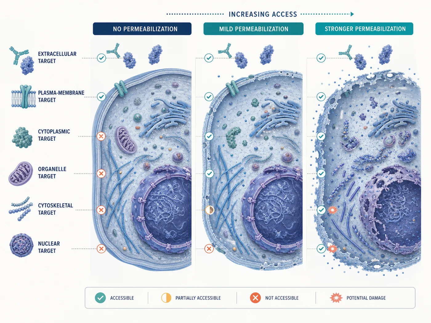
Start with the target location
The required access depends on the epitope, not only on the protein name. A membrane protein may expose one epitope to the extracellular space, another to the cytosol, and another to an organelle lumen.
| Epitope or target location | Minimum access needed | Structure at risk | Signs of excessive permeabilization |
|---|---|---|---|
| Extracellular cell-surface epitope | No detergent when fixation and antibody validation permit | Plasma-membrane continuity, receptor distribution, surface glycocalyx | New intracellular staining, loss of surface boundary, membrane collapse, or increased diffuse background |
| Cytosolic protein or cytosolic face of a membrane | Plasma-membrane access | Soluble protein pool, cortical cytoskeleton, membrane-associated complexes | Loss of soluble signal, cell shrinkage, disrupted edges, or altered membrane-associated pattern |
| Nuclear protein | Plasma membrane and nuclear-envelope access sufficient for the antibody and fixation state | Nuclear morphology, chromatin organization, nucleocytoplasmic boundaries | Collapsed nuclei, nonspecific nuclear haze, loss of compartment contrast, or broad extraction |
| Organelle-lumen epitope | Access across the plasma membrane and the relevant organelle membrane | Organelle membranes, lumenal contents, fine tubular or vesicular architecture | Fragmentation, disappearance of lumenal signal, loss of organelle shape, or conversion to diffuse staining |
| Cytoskeletal or weakly associated structure | Enough access for the antibody without extracting the structure | Filaments, focal adhesions, cortical networks, associated soluble factors | Broken filaments, punctate debris, loss of expected attachment sites, or condition-dependent redistribution |
Compare permeabilization methods
| Method | When useful | Main risks | What to check |
|---|---|---|---|
| No detergent after fixation | Surface-accessible epitopes or specimens already permeabilized by their preparation | Intracellular targets remain inaccessible; fixation may still alter the surface epitope | Known-positive surface signal, target-negative material, and evidence that intracellular markers remain excluded when exclusion is part of the design |
| Nonionic detergent after aldehyde fixation, commonly Triton X-100 | Broad access to cytoplasmic, nuclear, and many organelle-associated targets | Lipid extraction, membrane disruption, loss of soluble or weakly bound proteins, altered organelle morphology, and increased nonspecific access | Compare the lowest effective condition against no detergent or a milder condition while keeping fixation, antibody, and acquisition constant |
| Cold methanol as fixation plus permeabilization | Some cytoskeletal proteins and epitopes that perform poorly after aldehyde fixation | Lipid extraction, membrane and organelle distortion, shrinkage, and loss of soluble material | Treat it as a separate fixation route, not as a detergent strength within the same fixed specimen |
| Selective cholesterol-dependent agents such as digitonin or saponin | Specialized experiments that require differential access to the plasma membrane versus internal membranes | Selectivity varies with concentration, temperature, exposure time, cell type, membrane cholesterol, osmotic conditions, and prior fixation | Use accessible and protected compartment markers in the same experiment; do not infer selectivity from reagent name alone |
| Detergent included throughout blocking and antibody dilution | Maintaining access during long antibody incubations in a validated protocol | Long cumulative exposure can extract more material than a short dedicated permeabilization step | Record total detergent exposure and compare morphology and target retention with the complete staining workflow |
Concentration alone does not define severity. Reagent identity, temperature, duration, specimen geometry, fixation, agitation, and cumulative exposure all contribute to the final effect.
Consider the specimen type
| Specimen | Question | Keep in mind |
|---|---|---|
| Formaldehyde-fixed cultured cells | What is the least disruptive condition that exposes the required epitope? | Fixation and permeabilization can be optimized separately, but only one should change at a time. |
| Methanol-fixed cultured cells | Did the solvent route already provide adequate access while preserving the required structure? | Adding detergent may increase extraction without solving a fixation-dependent epitope problem. |
| Fresh-frozen cryosections | Did pre-fixation, post-section fixation, section thickness, or freezing already alter membrane access? | Sectioning exposes cut surfaces, but antibodies may still require controlled access through cells and internal membranes. |
| Previously fixed cryosections | Is poor access caused by cross-linking, section thickness, or inadequate permeabilization? | Stronger detergent cannot reverse every fixation-dependent masking effect. |
| FFPE sections | Are deparaffinization, rehydration, and retrieval adequate before considering detergent? | Permeabilization is not a substitute for complete paraffin removal or target-appropriate antigen retrieval. |
How to choose a permeabilization condition
- Identify the epitope location. Record whether the antibody recognizes an extracellular, cytosolic, nuclear, organelle-lumen, or unknown epitope.
- Start from the validated fixation method. Methanol fixation already permeabilizes; aldehyde fixation usually requires a separate permeabilization step for intracellular targets.
- Identify the structure that must remain intact. State whether the conclusion depends on plasma membrane, lipid droplets, ER, Golgi, mitochondria, lysosomes, nucleus, cytoskeleton, focal adhesions, or a soluble pool.
- Start with the mildest condition that gives access to the target. Use no detergent for an accessible surface epitope or one mild validated condition for an intracellular target.
- Use compartment markers when access is uncertain. Include one marker expected to be accessible and, when selective permeabilization is claimed, one marker expected to remain protected.
- Change one permeabilization variable at a time. Do not alter fixation, detergent identity, concentration, duration, antibody concentration, and imaging settings in the same comparison.
- Check that stronger permeabilization does not remove or distort the target. A brighter image can also result from increased background, extraction, collapse, or exposure of an off-target structure.
Compare permeabilization conditions
For an aldehyde-fixed cultured-cell assay, start by comparing no detergent with one low-detergent condition. Add a stronger condition only when the target remains inaccessible and morphology is still interpretable.
| Variable | Condition A | Condition B | Optional condition C | Keep constant |
|---|---|---|---|---|
| Access route | No detergent | One low, antibody-supported detergent condition | One stronger condition only when justified | Fixation, specimen state, reagent temperature, exchange method, and total staining time |
| Example fixed-cell screen | Buffer only | A short 0.05–0.1% Triton X-100 screen | A higher concentration or longer exposure drawn from a validated product protocol | Do not treat these values as universal optima |
| Labeling | Same primary clone and lot, concentration, secondary antibody, incubation, wash, counterstain, and mountant | Prepare dilutions together and stain in parallel | ||
| Acquisition | Same microscope, objective, optical configuration, exposure logic, gain, z-step, and processing | Acquire below saturation and retain raw channels | ||
Some published or manufacturer protocols use detergent continuously in blocking and antibody dilution rather than as a short separate step. Compare total detergent exposure, not concentration alone.
Surface and intracellular targets in the same experiment
When one marker is extracellular and another is intracellular, check the surface marker before adding a detergent condition that changes membrane access.
- Confirm whether the surface antibody requires live-cell labeling or can be used after fixation. Follow the antibody’s application-specific instructions and institutional biosafety procedures.
- Establish the surface marker alone without detergent. Confirm expected membrane localization and target-negative behavior.
- Test whether the planned fixation preserves the surface epitope. Fixation can reduce or redistribute surface staining even before detergent is added.
- Add permeabilization as a separate step. Check whether the surface pattern survives the detergent condition needed for the internal marker.
- Use order-specific omission controls. In multiplex indirect IF, confirm that secondary antibodies and later steps do not cross-detect the earlier surface-labeling layer.
- Inspect individual channels. Do not infer preservation from a merged image when the surface boundary has weakened or broadened.
Required controls
| Control | What it checks | What failure looks like |
|---|---|---|
| Known-positive specimen under every access condition | Can the complete fixation, access, staining, and acquisition workflow detect the expected pattern? | No signal cannot be distinguished from inadequate access or failed staining. |
| Target-negative biological or genetic material | Does the pattern depend on the intended target after permeabilization? | Stronger access reveals a new target-independent pattern. |
| Unstained specimen under every access condition | Did detergent, solvent, specimen damage, or mountant change channel background? | Background rises before antibody signal is considered. |
| Primary-omission or secondary-only control | Does broader access expose secondary-antibody, Fc-related, or detection-reagent binding? | Background increases as permeabilization becomes stronger. |
| Accessible compartment marker | Did the antibody reach the intended compartment? | The target and access marker both remain absent. |
| Protected compartment marker for selective access | Did internal membranes remain functionally inaccessible? | A supposedly protected lumenal marker becomes positive. |
| Morphology reference | Did the access condition preserve the structure required for interpretation? | Signal increases while cell or organelle geometry deteriorates. |
See the controls guide for target-dependence, detection-layer, autofluorescence, and multiplex control design.
Before choosing a permeabilization condition
- The antibody reaches the required compartment and produces the expected spatial pattern
- Target-negative material loses or substantially reduces the pattern
- Plasma membrane, organelle, nuclear, cytoskeletal, or soluble structures required by the question remain interpretable
- Unstained and detection-layer controls remain acceptable in every relevant channel
- The condition does not create new diffuse, edge-biased, punctate-debris, or compartment-independent staining
- Selective access is demonstrated with accessible and protected markers rather than assumed from reagent identity
- The result is reproducible across the required cells, sections, days, operators, and reagent lots
- The selected condition remains compatible with multiplexing, mounting, image acquisition, and quantitative analysis
Recognize permeabilization-related artifacts
| Observation | Possible explanation | Check |
|---|---|---|
| No intracellular signal but surface or nuclear counterstain is present | Insufficient access, fixation-dependent masking, wrong antibody application, or acquisition error | Known-positive specimen, accessible compartment marker, antibody validation route, and one controlled increase in access |
| Signal increases while cell boundaries disappear | Excessive lipid extraction or membrane disruption | Compare no detergent or a milder condition while keeping antibody and imaging constant |
| Organelle pattern becomes diffuse | Internal membrane disruption, lumenal-content loss, or extraction of membrane-associated proteins | Inspect an independent organelle marker and compare shorter or milder access |
| Cytoplasmic haze increases with detergent strength | Secondary background, nonspecific access, soluble-protein redistribution, or saturation | Secondary-only control, target-negative material, raw exposure values, and antibody titration |
| Nuclear staining appears only under the strongest condition | True access improvement, nuclear damage, or exposure of a target-independent nuclear binder | Known-positive and genetic-negative material, nuclear morphology, and a separate fixation comparison |
| Surface marker moves inward or becomes punctate | Membrane damage, receptor internalization before fixation, extraction, or cross-detection | Review labeling order, fixation delay, no-detergent surface control, and omission controls |
| Selective agent also reveals a lumenal marker | Over-permeabilization or loss of membrane selectivity | Reduce exposure and verify both cytosolic and lumenal control markers in the same cells |
Common permeabilization mistakes
| Mistake | Why it fails | Better approach |
|---|---|---|
| “Every intracellular target needs the same Triton condition.” | Compartment, fixation, membrane dependence, and extraction risk differ among targets. | Select access from epitope orientation and validate the lowest effective condition. |
| “More detergent means better penetration.” | Higher access may also mean greater extraction, structural collapse, and nonspecific binding. | Check target retention, morphology, target-negative material, and background as detergent exposure increases. |
| “Methanol fixation plus detergent is just a stronger version of formaldehyde plus detergent.” | Methanol changes fixation chemistry and extracts lipids before the detergent comparison begins. | Treat methanol as a separate fixation route and compare complete workflows. |
| “Digitonin or saponin is automatically plasma-membrane selective.” | Selectivity depends on membrane composition and experimental conditions. | Demonstrate access and protection with compartment markers in the same experiment. |
| “A bright nuclear pattern proves successful nuclear access.” | Nuclear damage, off-target binding, saturation, or autofluorescence can also create bright nuclei. | Use target-negative material, nuclear morphology, exposure control, and an access marker. |
| “Changing fixation and detergent together saves time.” | The cause of any improvement or artifact becomes uninterpretable. | Keep fixation constant first, then change one permeabilization variable at a time. |
What to record for permeabilization
- Specimen identity, preparation, fixation route, fixation duration, temperature, and delay to fixation
- Target name, antibody clone and lot, epitope or domain when known, expected compartment, and application-specific validation
- Structure that must remain intact and the morphology marker used to evaluate it
- Permeabilizing reagent, supplier or grade, stock preparation, concentration, buffer, temperature, duration, volume, and agitation
- Whether detergent remained in blocking, primary, secondary, or wash buffers and the total exposure time
- No-detergent, mild, stronger, or selective conditions compared and the reason each was included
- Known-positive, target-negative, unstained, secondary-only, accessible-compartment, protected-compartment, and morphology controls used
- Observed target pattern, background, membrane integrity, organelle morphology, nuclear morphology, extraction, and sample loss
- Microscope, objective, channels, exposure, gain, z-step, saturation rule, processing, and raw-file location
- Acceptance decision, rejected conditions, repeat count, lot changes, and revalidation trigger
References and protocol sources
- Immunofluorescence protocol for cell-based imaging Manufacturer protocol
Cell Signaling Technology
Product-specific validation and formaldehyde-versus-methanol sample preparation.
- Immunofluorescent staining of intracellular antigens on cultured cells Manufacturer protocol
Thermo Fisher Scientific
Cultured-cell fixation, washing, blocking, antibody incubation, mounting, and storage starting conditions.
- Immunofluorescence protocol for frozen tissue (IF-F) Manufacturer protocol
Cell Signaling Technology
Charged slides, cryosection thickness, fixation, blocking, antibody incubation, washing, and mounting starting conditions.
- Overview of immunofluorescence techniques Technical guide
Cell Signaling Technology
Matching antibodies and protocols to biological material and sample processing.
- Immunofluorescence staining Methods paper
Julie G. Donaldson . Current Protocols in Cell Biology (2015) . DOI: 10.1002/0471143030.cb0403s69
Indirect immunofluorescence workflow, controls, fixation, permeabilization, and specimen handling.
-
S. R. Yang, B. K. Maity, and S. Chong . The Journal of Physical Chemistry B (2023) . DOI: 10.1021/acs.jpcb.3c01658
Fixation-dependent redistribution, cross-linking and organic-solvent trade-offs, and interpretation limits.
- High-quality immunofluorescence of cultured cells Methods paper
Dibyendu Bhattacharyya, Adam T. Hammond, and Benjamin S. Glick . Methods in Molecular Biology (2010) . DOI: 10.1007/978-1-60327-412-8_24
A specialized cultured-cell method illustrating why delicate structures can require protocol-specific preservation and validation.