Scope
This guide is primarily for formalin-fixed paraffin-embedded tissue used in immunofluorescence. It also covers the narrower situation in which an aldehyde-fixed frozen section has antibody-specific evidence supporting retrieval.
Antigen retrieval can improve access to epitopes altered or masked by fixation and processing. It cannot restore tissue that was inadequately fixed, overprocessed, incompletely deparaffinized, detached from the slide, dried, extracted, or structurally destroyed.
First decide whether retrieval is needed
| Specimen | Typical use | Check first |
|---|---|---|
| FFPE section | Retrieval is commonly required, but the route is antibody- and tissue-dependent. | Complete paraffin removal, graded rehydration, adequate slide adhesion, fixation history, and FFPE application evidence for the antibody. |
| Aldehyde-fixed frozen section | Optional branch, not a routine requirement. | Antibody-specific evidence or a controlled comparison showing that retrieval improves target-dependent signal without unacceptable section loss or morphology damage. |
| Alcohol- or acetone-fixed frozen section | Usually no retrieval unless a validated method explicitly requires it. | Whether the fixation route already exposes the epitope and whether heating or enzymes would add damage rather than useful access. |
| Fixed cultured cells | Usually optimize fixation and permeabilization first. | Application-specific instructions demonstrating that retrieval is necessary for this antibody, cell preparation, and target. |
| Unfixed or live-cell labeling | Conventional HIER or PIER does not belong in the workflow. | Use a method designed for live-cell or unfixed material. |
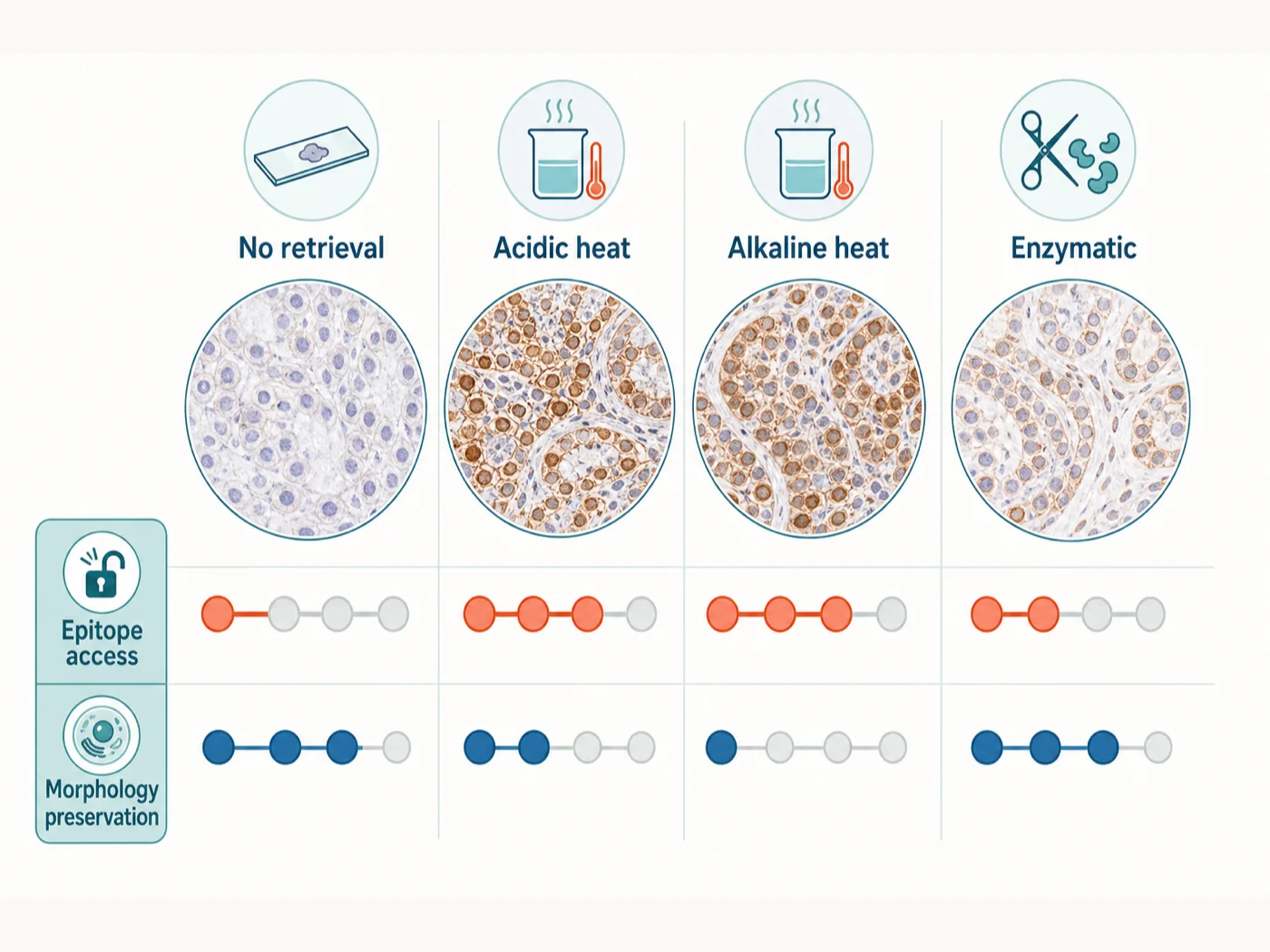
Compare retrieval methods
| Method | When used | Main risks | When to stop |
|---|---|---|---|
| Validated no retrieval | Antibodies and epitopes that remain accessible after the documented fixation and processing route. | A true target may remain masked, producing a false-negative or weak result. | Accept only when positive material is detectable, negative material behaves appropriately, morphology is intact, and the result is reproducible. |
| Heat-induced epitope retrieval (HIER) | Common first retrieval route for many formalin-fixed, paraffin-embedded targets. | Tissue detachment, folds, cracking, altered morphology, increased background or autofluorescence, buffer evaporation, and condition-dependent loss of some epitopes. | Stop escalation when signal gains are no longer target-dependent or when tissue retention, morphology, or background becomes unacceptable. |
| Proteolytic enzyme-induced epitope retrieval (PIER) | Selected targets or tissues for which validated protease digestion performs better than heat. | Overdigestion, loss of tissue architecture, cleavage of the target or other proteins, section lifting, and difficult run-to-run control. | Stop digestion at the validated time, quench or wash exactly as specified, and reject conditions with morphology loss or new target-independent staining. |
| Combined or sequential HIER and PIER | Specialized workflows supported by strong product-specific or published evidence. | Compounded tissue damage and an optimization space too large to interpret reliably. | Do not use as the first screen. Introduce only after each component route has been evaluated independently. |
Record HIER temperature and time, not only the buffer
“Citrate retrieval” or “high-pH retrieval” does not fully describe the condition. Record the buffer, actual temperature, heating time, device, and cooling history experienced by the section.
| Variable | Why it matters | What to record |
|---|---|---|
| Buffer identity and pH | Low-pH citrate, neutral or EDTA-containing buffers, and alkaline Tris-EDTA conditions can expose different epitopes and alter tissue morphology differently. | Reagent name, formulation, lot, preparation date, measured pH, and whether a proprietary concentrate was diluted correctly. |
| Actual solution temperature | The instrument setpoint does not guarantee the temperature experienced by the slide. | Device, preheating method, measured or validated solution temperature, ramp time, and whether slides were introduced before or after heating. |
| Time at retrieval temperature | Temperature and duration act together; a longer exposure is not interchangeable with a shorter exposure at a different temperature. | Start and stop definition, actual high-temperature interval, and any interruption or reheating. |
| Container, volume, and slide load | Geometry and load change heating rate, evaporation, mixing, and temperature uniformity. | Container type, buffer volume, number and orientation of slides, lid or seal, and evaporation control. |
| Submersion and drying prevention | Exposed tissue edges or local evaporation can create irreversible edge artifacts and section loss. | Whether slides remained fully submerged and whether bubbles or low buffer volume were observed. |
| Cooling phase | Controlled cooling is part of the protocol and influences handling, tissue stress, and reproducibility. | Cooling in retrieval solution or transfer buffer, duration, target temperature, and agitation. |
A current Thermo Fisher high-pH FFPE protocol uses fully submerged slides at approximately 98°C for 20 minutes followed by about 20 minutes of cooling in the retrieval solution. Treat this as a defined product workflow and comparison point, not a universal optimum.
PIER requires controlled digestion
Use PIER when the antibody and tissue support a proteolytic method, not simply because HIER produced weak signal. Protease activity varies with reagent, lot, concentration, buffer, pH, temperature, tissue fixation, and time.
| Variable | Risk if uncontrolled | What to record |
|---|---|---|
| Enzyme identity and activity | Trypsin, proteinase K, pepsin, and other enzymes do not cleave the same substrates or behave interchangeably. | Enzyme, supplier, catalog number, lot, activity specification, preparation, and storage. |
| Concentration and buffer | Incorrect dilution or incompatible pH changes digestion rate and target retention. | Final concentration, buffer composition, pH, calcium or other cofactors, and preparation time. |
| Temperature and duration | Small increases can shift the result from inadequate retrieval to tissue destruction. | Validated temperature, actual incubation time, slide equilibration, and timing accuracy. |
| Stop or wash step | Digestion can continue during handling if the enzyme is not removed or inactivated promptly. | Exact stop method, wash composition, wash count, and elapsed time before blocking. |
| Morphology checkpoint | Strong signal can conceal overdigestion, section thinning, and loss of diagnostic architecture. | Brightfield or transmitted-light morphology before and after digestion, tissue retention, and predefined rejection criteria. |
A current Thermo Fisher fluorescent FFPE trypsin workflow provides 0.1% trypsin at 37°C for approximately 10–20 minutes as a product-specific starting condition. Enzyme conditions must still be optimized for the tissue, fixation history, and antibody.
How to choose a retrieval method
- Confirm the specimen type. Determine whether the sample is FFPE, aldehyde-fixed frozen tissue, alcohol-fixed frozen tissue, or another preparation before adding retrieval.
- Check the steps before retrieval. For FFPE, verify deparaffinization, rehydration, slide adhesion, section quality, fixation duration, and processing history.
- Check antibody instructions for the same application. Use the antibody’s FFPE, frozen-tissue, or other application-specific preparation as the first candidate, including whether retrieval is omitted, heat-based, or enzymatic.
- Compare one retrieval method at a time. Start with no retrieval versus one HIER condition, or no retrieval versus one validated PIER condition. Do not vary HIER, PIER, pH, time, and antibody concentration together.
- Decide which tissue structures must remain intact. Note the tissue architecture, membrane boundary, nuclear feature, extracellular matrix, or cell type required for interpretation.
- Choose the controls and rejection conditions before starting. Include positive, target-negative, unstained, and detection-layer controls before examining experimental sections.
- Acquire controls first. Check target detectability separately from autofluorescence, secondary background, tissue loss, and exposure saturation.
Compare retrieval conditions
Start with no retrieval and one antibody-supported retrieval condition. Add a second pH or PIER condition only if the first comparison does not answer whether retrieval improves staining without unacceptable tissue damage.
| Stage | Condition A | Condition B | Optional condition C | Keep constant |
|---|---|---|---|---|
| Retrieval route | Validated no retrieval | One antibody-supported HIER condition | One alternate HIER pH or one validated PIER condition | Block, section thickness, slide type, deparaffinization, rehydration, and tissue batch |
| HIER variables | Not applicable | One defined buffer, temperature, duration, device, and cooling sequence | Change only one major retrieval variable | Do not alter pH and thermal exposure simultaneously unless reproducing a defined complete protocol |
| Labeling | Same primary clone and lot, concentration, secondary or direct conjugate, incubation, wash, counterstain, and mountant | Prepare reagents together and stain in parallel | ||
| Acquisition | Same microscope, objective, optical configuration, exposure logic, gain, z-step, saturation rule, and processing | Retain raw individual channels | ||
Required controls
| Control | What it checks | What failure looks like |
|---|---|---|
| Known-positive tissue under every retrieval condition | Can this complete preparation and staining workflow reveal the expected pattern? | Experimental absence cannot be distinguished from retrieval or staining failure. |
| Target-negative biological or genetic material | Does the retrieved pattern remain dependent on the intended target? | A stronger retrieval condition creates a new target-independent structure or diffuse pattern. |
| Unstained serial section under every condition | Did heat, enzyme treatment, fixation, pigment, or mountant increase fluorescence in any channel? | Apparent signal rises before antibody labeling is considered. |
| Primary-omission or secondary-only control | Did retrieval expose new binding sites for the fluorescent secondary or downstream detection layer? | Background increases after retrieval even without the intended primary antibody. |
| No-retrieval comparison | Did the retrieval condition provide a measurable benefit over the same tissue without retrieval? | The target already stains adequately without retrieval or the retrieved condition only adds background and damage. |
| Morphology and tissue-retention reference | Did the section remain attached and structurally interpretable? | Signal increases while folds, cracks, lifting, digestion, or architectural loss make the result unreliable. |
| Single-color control for multiplex work | Did retrieval or increased brightness alter channel contamination? | A fluorophore appears in a neighboring channel after exposure is increased. |
See the controls guide for target-dependence, detection-layer, autofluorescence, and multiplex control design.
Before choosing a retrieval condition
- Known-positive material shows the expected cellular, tissue, and subcellular distribution below detector saturation
- Target-negative material loses or substantially reduces the pattern without a new retrieval-dependent off-target pattern
- Unstained and detection-layer controls remain acceptable in every relevant channel
- Tissue remains attached, hydrated, and free of retrieval-induced folds, cracks, edge lifting, or major section loss
- Nuclear, membrane, extracellular-matrix, and tissue architecture required by the biological question remain interpretable
- The selected condition performs better than no retrieval or the alternative route on the complete evidence, not only on peak brightness
- The result is reproducible across the required blocks, sections, days, operators, devices, buffer lots, and antibody lots
- The condition remains compatible with multiplexing, fluorescence background, mounting, acquisition, and quantitative analysis
Recognize retrieval-related artifacts
| Observation | Possible explanation | Check |
|---|---|---|
| No signal in positive tissue | Wrong retrieval route, inadequate thermal exposure, enzyme underdigestion, failed staining, incomplete deparaffinization, or acquisition error | Verify the positive control, paraffin removal, actual temperature, antibody application, channel, and one controlled route change |
| Signal rises with diffuse or extracellular haze | Excessive retrieval, secondary background, tissue damage, or exposure saturation | Target-negative, secondary-only, unstained controls, raw exposure values, and a milder condition |
| Section lifts after heating | Inadequate slide adhesion, old or poorly dried section, severe buffer or thermal stress, abrupt pressure change, or forceful washing | Review slide type, section age, baking, device cycle, cooling, tissue composition, and edge lifting before antibody staining |
| Tissue looks swollen, cracked, or fragmented | Excess heat, alkaline exposure, rapid thermal change, evaporation, or overdigestion | Inspect transmitted-light morphology, actual temperature, buffer volume, cooling, and a shorter or milder route |
| Strong background in the unstained section | Intrinsic fluorescence, formalin-associated fluorescence, pigment, heat-related change, or mountant contribution | Acquire unstained controls by channel before raising antibody concentration or adding amplification |
| One cell type disappears while another brightens | Differential tissue damage, epitope destruction, extraction, or a route favoring one target compartment | Compare serial sections, morphology markers, target-negative material, and an alternate route without changing the antibody concentration |
| PIER gives strong signal with blurred architecture | Overdigestion or continuing enzyme activity during handling | Shorten exposure, verify temperature and stop step, and inspect tissue before antibody incubation |
Common retrieval mistakes
| Mistake | Why it fails | Better approach |
|---|---|---|
| “Every FFPE antibody needs high-pH HIER.” | Epitopes, fixation histories, tissues, and antibodies respond differently. | Start from application-specific evidence and compare a limited set including no retrieval when justified. |
| “The strongest signal is the best retrieval.” | Brightness can rise because of background, tissue damage, off-target access, or saturation. | Use positive, negative, unstained, secondary-only, morphology, and tissue-retention evidence together. |
| “Longer heating is equivalent to higher temperature.” | Thermal history, buffer chemistry, device geometry, and cooling interact nonlinearly. | Record and validate the complete condition rather than substituting one variable casually. |
| “PIER is simply a stronger backup when HIER fails.” | Proteases can destroy the target and architecture while revealing unrelated sites. | Use PIER only with target- and tissue-specific justification and a controlled digestion range. |
| “Retrieval can rescue incomplete deparaffinization.” | Residual paraffin prevents reproducible wetting and reagent access. | Correct clearing and rehydration before evaluating retrieval. |
| “Frozen sections should always receive citrate retrieval.” | Many frozen workflows do not require retrieval, and heating can detach or damage sections. | Use retrieval only when aldehyde fixation and antibody-specific evidence justify the branch. |
| “Changing retrieval, antibody concentration, and exposure together saves time.” | The cause of improvement or artifact becomes uninterpretable. | Keep labeling and acquisition constant while changing one retrieval condition at a time. |
What to record for antigen retrieval
- Tissue identity, block, biological replicate, cold ischemia, fixative, fixation duration, processor program, block age, and storage
- Section thickness, slide type or coating, section date, baking or drying conditions, folds, compression, and pre-retrieval adhesion
- Clearing reagent, deparaffinization sequence, graded rehydration, and confirmation that tissue remained hydrated
- Retrieval route: none, HIER, PIER, or validated combined sequence, with the reason for selection
- HIER buffer, formulation, lot, preparation, pH, device, container, volume, slide load, preheating, actual temperature, duration, evaporation control, and cooling
- PIER enzyme, supplier, catalog number, lot, activity, concentration, buffer, pH, temperature, duration, and stop method
- Tissue retention and morphology before retrieval, after retrieval, after washing, and after staining
- Primary and detection reagents with clone, lot, concentration, dilution, incubation, wash, fluorophore, and amplification details
- Known-positive, target-negative, unstained, secondary-only, no-retrieval, morphology, and single-color controls used
- Microscope, objective, channels, exposure, gain, z-step, saturation rule, processing, raw-file location, acceptance decision, repeat count, and revalidation trigger
References and protocol sources
- Paraffin removal and antigen retrieval Technical guide
Thermo Fisher Scientific
Deparaffinization, rehydration, and epitope retrieval as distinct preparation stages.
- FFPE tissue high-pH antigen retrieval, direct fluorescent method Manufacturer protocol
Thermo Fisher Scientific
Slide warming, deparaffinization, graded rehydration, high-pH HIER, cooling, staining, and storage.
- FFPE tissue trypsin digestion antigen retrieval, indirect fluorescent method Manufacturer protocol
Thermo Fisher Scientific
Proteolytic retrieval starting conditions and indirect fluorescent detection workflow.
- IHC antigen retrieval protocol Manufacturer protocol
Abcam
Heat-induced and enzymatic retrieval as target- and tissue-dependent strategies.
- Frozen tissues with citrate antigen retrieval Manufacturer protocol
Cell Signaling Technology
Antigen retrieval as an optional antibody-specific branch for frozen sections.
-
Cell Signaling Technology
FFPE application validation, formalin-associated autofluorescence, sensitivity limits, and amplification considerations.
- Controls for immunohistochemistry: The Histochemical Society’s standards of practice for validation of immunohistochemical assays Consensus guideline
Stephen M. Hewitt, Denis G. Baskin, Charles W. Frevert, William L. Stahl, and Eduardo Rosa-Molinar . Journal of Histochemistry & Cytochemistry (2014) . DOI: 10.1369/0022155414545224
Positive and negative control requirements, limits of primary-omission and absorption controls, and application-specific evidence for antibody specificity.