Scope
This guide addresses a specific fixed-cell immunofluorescence question: whether 4% paraformaldehyde and 0.1% Triton X-100 can be applied simultaneously for fixation and permeabilization. It is intended for adherent cultured cells and does not establish a universal condition for tissue sections, FFPE material, live-cell labeling, or every antibody.
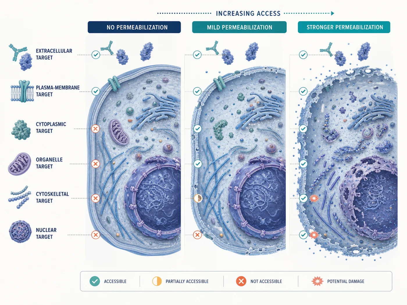
Short answer
A combined paraformaldehyde-and-detergent step can be a valid protocol-specific method, but it is not interchangeable with sequential fixation followed by permeabilization. During the combined step, cross-linking, membrane extraction, solute movement, and access changes occur at the same time. Compare the combined method with sequential fixation and permeabilization, and check whether target localization and cell morphology remain acceptable.
The numbers “4% paraformaldehyde” and “0.1% Triton X-100” describe only two concentrations. Temperature, exposure time, buffer composition, cell state, solution exchange, target mobility, and total detergent exposure can materially change the result.
Why simultaneous and sequential methods are not equivalent
| Variable | Sequential fixation then permeabilization | Simultaneous fixation and permeabilization |
|---|---|---|
| Order of preservation and access | Cross-linking begins before a dedicated membrane-access step | Cross-linking and detergent-mediated access begin together |
| Soluble or weakly associated material | Some material may be immobilized before detergent exposure | Material can move or be extracted while fixation is still developing |
| Membrane-dependent morphology | Membranes are challenged after the selected fixation interval | Membrane extraction begins during fixation and may alter organelles or boundaries earlier |
| Experimental interpretation | Fixation and access can be varied as separate factors | A brighter or weaker result cannot be attributed to fixation or permeabilization alone |
| Transferability | Often easier to compare with conventional antibody protocols | Must be tested for the specific target, cell type, and complete staining method |
When a combined method is worth testing
- An antibody or published method explicitly uses a combined aldehyde-and-detergent step for the same application and comparable specimen.
- The biological structure is poorly preserved or inaccessible under a conventional sequential method, and there is a specific reason to test detergent exposure during fixation.
- The experiment can include sequential and combined conditions in parallel rather than replacing the established method without comparison.
- The conclusion can be checked with target-positive and target-negative material, morphology references, and raw individual channels.
Compare simultaneous and sequential treatment
| Condition | Purpose | Keep constant |
|---|---|---|
| Sequential reference | Use the current validated fixation step followed by the current validated permeabilization step | Cell preparation, antibody lot and concentration, incubation, washes, mountant, microscope, exposure logic, and processing |
| Combined test | Test the proposed paraformaldehyde-and-Triton method as one complete treatment | Use matched cultures and stain in parallel with the sequential reference |
| Optional fixation-only comparison | Determine whether the target is surface-accessible or whether fixation alone creates the observed pattern | Do not add this condition when the target cannot plausibly be detected without intracellular access |
| Optional milder-access comparison | Determine whether a lower cumulative detergent exposure preserves morphology while maintaining adequate access | Change only one access variable from the selected reference method |
- Use matched specimens. Seed, treat, and process cultures together so cell state is not confounded with preparation method.
- Record the actual solution history. Include concentrations, buffer, pH, temperature, exposure time, exchange method, and wash timing.
- Acquire controls before choosing display settings. Establish background and the usable detector range from raw channels.
- Compare localization, not brightness alone. Inspect cell boundaries, organelles, nuclei, soluble pools, expected positive structures, and target-negative regions.
- Repeat the experiment before adopting the method. One good field is not enough to establish reproducibility.
Minimum controls
| Control | What it checks |
|---|---|
| Known-positive specimen under both methods | Can each complete preparation and staining method detect the expected target pattern? |
| Target-negative biological or genetic material | Does the pattern remain target-dependent after the combined exposure? |
| Unstained specimen under both methods | Does the combined condition change intrinsic fluorescence, debris, morphology, or mountant-related background? |
| Primary-omission or secondary-only control | Does broader or earlier access increase detection-reagent binding? |
| Structural or compartment reference | Are membranes, nuclei, organelles, cytoskeleton, or other structures required for interpretation preserved comparably? |
What to check before adopting the combined method
- The expected target localization is retained and remains target-dependent
- Known-positive material is detected without detector saturation
- Target-negative and detection-reagent controls remain within the predefined background limit
- Cell, nuclear, membrane, organelle, or cytoskeletal morphology required for the conclusion remains interpretable
- The combined method does not create a new diffuse, punctate, edge, or compartment pattern unsupported by controls
- The result is reproducible across the required cultures, fields, days, and operators
- The reason for choosing the combined method over the sequential reference is documented
If the result looks wrong, check these first
| Observation | Possible explanation | Check next |
|---|---|---|
| Combined method is brighter but more diffuse | Improved access, extraction, redistribution, or increased nonspecific binding | Inspect target-negative, omission, morphology, and sequential-reference channels under identical acquisition |
| Membrane or organelle boundaries disappear | Detergent exposure is incompatible with the structure required for interpretation | Return to the sequential method or compare one lower cumulative detergent condition |
| Soluble or weakly associated target is lost | Extraction may occur before sufficient immobilization | Compare sequential fixation before detergent while holding staining and imaging constant |
| Nuclear signal improves but cytoplasmic structure worsens | The combined method solves one access problem while creating a preservation problem | Define which compartment is essential and compare a milder or shorter detergent exposure |
| Only one antibody benefits | The result is antibody- or epitope-specific rather than a general laboratory condition | Do not apply the condition to other antibodies or the whole panel without testing them |
What to record
- Cell line, passage or culture state, substrate, confluence, treatment, and sampling time
- Paraformaldehyde source and preparation, Triton X-100 source, buffer, pH, concentrations, temperature, and exposure time
- Whether solutions were pre-mixed, added sequentially without washing, or exchanged as a single combined reagent
- Wash buffer, volume, number, duration, temperature, and agitation
- Primary and secondary antibody supplier, catalog number, clone, lot, concentration, incubation, and application evidence
- Positive, negative, unstained, omission, structural-reference, and sequential-workflow controls
- Microscope, objective, channels, exposure, gain, z-step, saturation rule, raw-file location, and processing
- Final decision, excluded fields, deviations, repeat result, and reason for retaining or rejecting the combined method
References and protocol sources
- Immunofluorescence staining Methods paper
Julie G. Donaldson . Current Protocols in Cell Biology (2015) . DOI: 10.1002/0471143030.cb0403s69
Indirect immunofluorescence workflow, controls, fixation, permeabilization, and specimen handling.
-
S. R. Yang, B. K. Maity, and S. Chong . The Journal of Physical Chemistry B (2023) . DOI: 10.1021/acs.jpcb.3c01658
Fixation-dependent redistribution, cross-linking and organic-solvent trade-offs, and interpretation limits.
- High-quality immunofluorescence of cultured cells Methods paper
Dibyendu Bhattacharyya, Adam T. Hammond, and Benjamin S. Glick . Methods in Molecular Biology (2010) . DOI: 10.1007/978-1-60327-412-8_24
A specialized cultured-cell method illustrating why delicate structures can require protocol-specific preservation and validation.
- Immunofluorescence protocol for cell-based imaging Manufacturer protocol
Cell Signaling Technology
Product-specific validation and formaldehyde-versus-methanol sample preparation.
- Immunofluorescent staining of intracellular antigens on cultured cells Manufacturer protocol
Thermo Fisher Scientific
Cultured-cell fixation, washing, blocking, antibody incubation, mounting, and storage starting conditions.